44 diagram of a labeled microscope
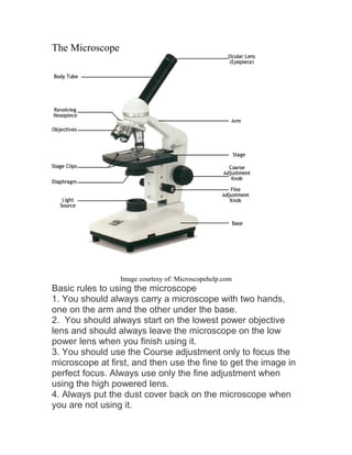
Microscope labeled diagram - SlideShare Microscope labeled diagram Oct. 30, 2013 • 6 likes • 27,530 views Download Now Download to read offline Pisgah High School Follow 1. The Microscope Image courtesy of: Microscopehelp.com Basic rules to using the microscope 1. You should always carry a microscope with two hands, one on the arm and the other under the base. 2. Microscope Types (with labeled diagrams) and Functions Simple microscope labeled diagram Simple microscope functions It is used in industrial applications like: Watchmakers to assemble watches Cloth industry to count the number of threads or fibers in a cloth Jewelers to examine the finer parts of jewelry Miniature artists to examine and build their work Also used to inspect finer details on products
Label the microscope — Science Learning Hub In this interactive, you can label the different parts of a microscope. Use this with the Microscope parts activity to help students identify and label the main parts of a microscope and then describe their functions. Drag and drop the text labels onto the microscope diagram.
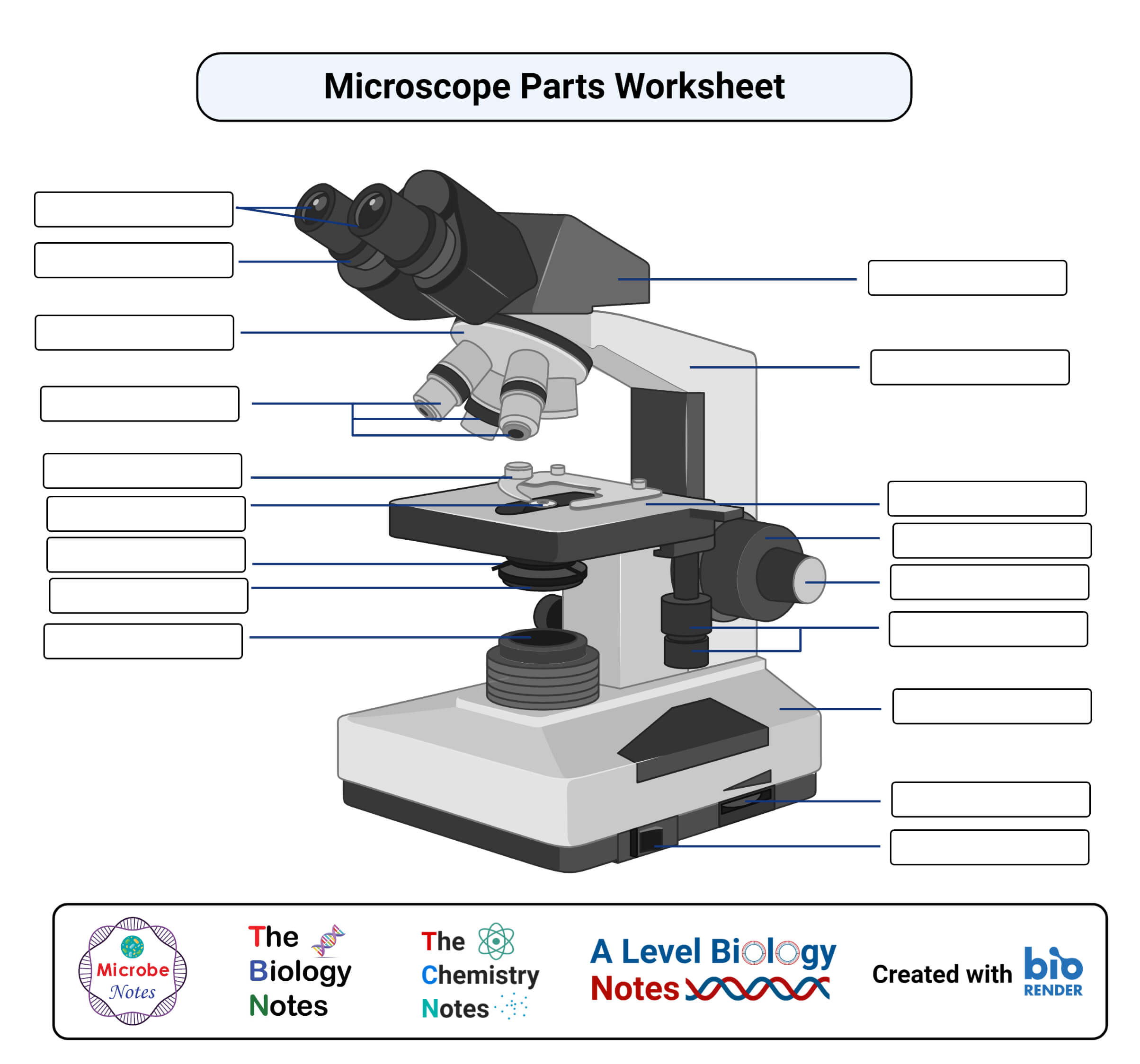
Diagram of a labeled microscope
Microscope Parts, Function, & Labeled Diagram - slidingmotion Microscope parts labeled diagram gives us all the information about its parts and their position in the microscope. Microscope Parts Labeled Diagram The principle of the Microscope gives you an exact reason to use it. It works on the 3 principles. Magnification Resolving Power Numerical Aperture. Parts of Microscope Head Base Arm Eyepiece Lens Compound Microscope Parts, Functions, and Labeled Diagram Compound Microscope Parts, Functions, and Labeled Diagram Parts of a Compound Microscope Each part of the compound microscope serves its own unique function, with each being important to the function of the scope as a whole. Labelled Diagram of Compound Microscope - Biology Discussion The below mentioned article provides a labelled diagram of compound microscope. Part # 1. The Stand: The stand is made up of a heavy foot which carries a curved inclinable limb or arm bearing the body tube. The foot is generally horse shoe-shaped structure (Fig. 2) which rests on table top or any other surface on which the microscope in kept.
Diagram of a labeled microscope. Label the Microscope Diagram | Download Scientific Diagram Download scientific diagram | Label the Microscope Diagram from publication: Laboratory Exercises in Microbiology: Discovering the Unseen World through Hands-on Investigation | Microbiology ... Neuron under Microscope with Labeled Diagram - AnatomyLearner Here, you will also find the diagrams of different neuron types under a microscope. The neuron diagram shows the different parts (axon, dendrites, and cell body) of the neurons. You may also find more neuron diagrams on social media of anatomy learners. Frequently asked questions on a neuron under microscope Label a Microscope - Storyboard That Knowing the names of the different parts of the microscope is essential to be able to use one properly. Create a poster that labels the parts of a microscope and includes descriptions of what each part does. Click "Start Assignment". Use a landscape poster layout (large or small). Search for a diagram of a microscope. PDF Label parts of the Microscope: Answers Label parts of the Microscope: Answers Coarse Focus Fine Focus Eyepiece Arm Rack Stop Stage Clip . Created Date: 20150715115425Z ...
Compound Microscope Labeled Diagram | Quizlet QUESTION. The total magnification of a specimen being viewed with a 10X ocular lens and a 40X objective lens is. 15 answers. QUESTION. a mosquito beats its wings up and down 600 times per second, which you hear as a very annoying 600 Hz sound. if the air outside is 20 C, how far would a sound wave travel between wing beats. 2 answers. Microscope Labeling - The Biology Corner Students label the parts of the microscope in this photo of a basic laboratory light microscope. Can be used for practice or as a quiz. ... 20. A microscope has an ocular objective of 10x and a high power objective of 50x, what is the microscope's total magnification? _____ Parts of Stereo Microscope (Dissecting microscope) - labeled diagram ... Labeled part diagram of a stereo microscope Major structural parts of a stereo microscope There are three major structural parts of a stereo microscope. The viewing Head includes the upper part of the microscope, which houses the most critical optical components, including the eyepiece, objective lens, and light source of the microscope. Microscope Diagram Labeled, Unlabeled and Blank | Parts of a Microscope ... Print a microscope diagram, microscope worksheet, or practice microscope quiz in order to learn all the parts of a microscope. These parts of a microscope printables include word searches, crossword puzzles, and vocabulary worksheets. A diagram showing all of the parts of a compound light microscope. Help students learn and remember the parts ...
Compound Microscope- Definition, Labeled Diagram, Principle, Parts, Uses The optical microscope often referred to as the light microscope, is a type of microscope that uses visible light and a system of lenses to magnify images of small subjects. There are two basic types of optical microscopes: Simple microscopes. Compound microscopes. The term "compound" in compound microscopes refers to the microscope having ... A Study of the Microscope and its Functions With a Labeled Diagram A Study of the Microscope and its Functions With a Labeled Diagram To better understand the structure and function of a microscope, we need to take a look at the labeled microscope diagrams of the compound and electron microscope. These diagrams clearly explain the functioning of the microscopes along with their respective parts. M mooketsi Microscope, Microscope Parts, Labeled Diagram, and Functions Microscope, Microscope Parts, Labeled Diagram, and Functions What is Microscope? A microscope is a laboratory instrument used to examine objects that are too small to be seen by the naked eye. It is derived from Ancient Greek words and composed of mikrós, "small" and skopeîn,"to look" or "see". Labeled Microscope and Basics of Life Diagram | Quizlet the lens at the top that you look through. They are usually 10X or 15X power. Base The bottom of the microscope, used for support and carrying Body tube Connects the eyepiece to the objective lenses Coarse adjustment Brings the specimen into general focus. Fine adjustment Moves the stage slightly to sharpen the image Objective lens
Parts of a microscope with functions and labeled diagram
Simple Microscope - Parts, Functions, Diagram and Labelling Parts of the optical parts are as follows: Mirror - A simple microscope has a plano-convex mirror and its primary function is to focus the surrounding light on the object being examined. Lens - The biconvex lens is placed above the stage and its function is to magnify the size of the object being examined.

Solved and In the following microscope diagram, the parts ...
Simple Microscope - Diagram (Parts labelled), Principle, Formula and Uses Parts of a Simple Microscope A simple microscope consists of Optical parts Mechanical parts Labeled Diagram of simple microscope parts Optical parts The optical parts of a simple microscope include Lens Mirror Eyepiece Lens A simple microscope uses biconvex lens to magnify the image of a specimen under focus.

Microscope Labeling
Compound Microscope Parts - Labeled Diagram and their Functions - Rs ... Labeled diagram of a compound microscope Major structural parts of a compound microscope There are three major structural parts of a compound microscope. The head includes the upper part of the microscope, which houses the most critical optical components, and the eyepiece tube of the microscope.
Compound Microscope Parts, Functions, and Labeled Diagram ...
Microscope Labeling - The Biology Corner Microscope Labeling. This simple worksheet pairs with a lesson on the light microscope, where beginning biology students learn the parts of the light microscope and the steps needed to focus a slide under high power. The labeling worksheet could be used as a quiz or as part of direct instruction where students label the microscope as you go ...

Labeling the Parts of the Microscope | Microscope World Resources
Microscope Parts and Functions With Labeled Diagram and Functions How ... Most specimens are mounted on slides, flat rectangles of thin glass. The specimen is placed on the glass and a cover slip is placed over the specimen. This allows the slide to be easily inserted or removed from the microscope. It also allows the specimen to be labeled, transported, and stored without damage.

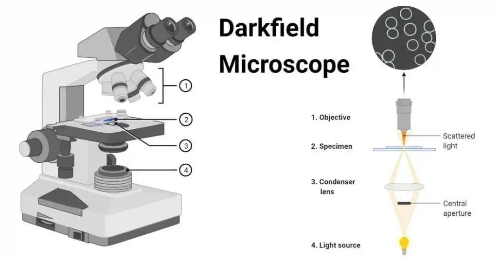
Types of Microscopes: Definition, Working Principle, Diagram ...
Blood Histology Slides with Description and Labeled Diagram The blood is a specialized connective tissue that is fluid and circulates through the vascular channel. In the blood histology slide, you will find different types of cells with their specific features. This might be a short article where I will show you all the cells from the blood microscope slide with a labeled diagram and actual pictures.

Simple Microscope - Parts, Functions, Diagram and Labelling ...
22 Parts Of a Microscope With Their Function And Labeled Diagram The field diaphragm control is located around the lens located in the base. Hinge Screw -This screw fixes the arm to the base and allow for the tilting of the arm. Stage Clips - They hold the slide firmly onto the stage. On/OFF Switch - This switch on the base of the microscope turns the illuminator off and on.

Easy labeled diagram of Microscope - YouTube
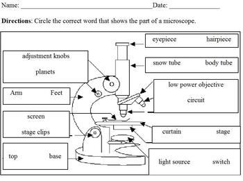
Labeling the Parts of the Microscope Labeling the Parts of the Microscope This activity has been designed for use in homes and schools. Each microscope layout (both blank and the version with answers) are available as PDF downloads. You can view a more in-depth review of each part of the microscope here. Download the Label the Parts of the Microscope PDF printable version here.

10+ Free Microscopy & Microscope Illustrations
A Study of the Microscope and its Functions With a Labeled Diagram These labeled microscope diagrams and the functions of its various parts, attempt to simplify the microscope for you. However, as the saying goes, 'practice makes perfect', here is a blank compound microscope diagram and blank electron microscope diagram to label. Download the diagrams and practice labeling the different parts of these ...
Microscope Types (with labeled diagrams) and Functions
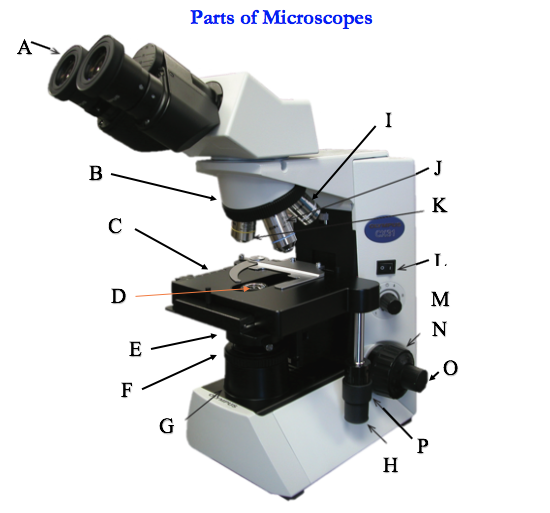
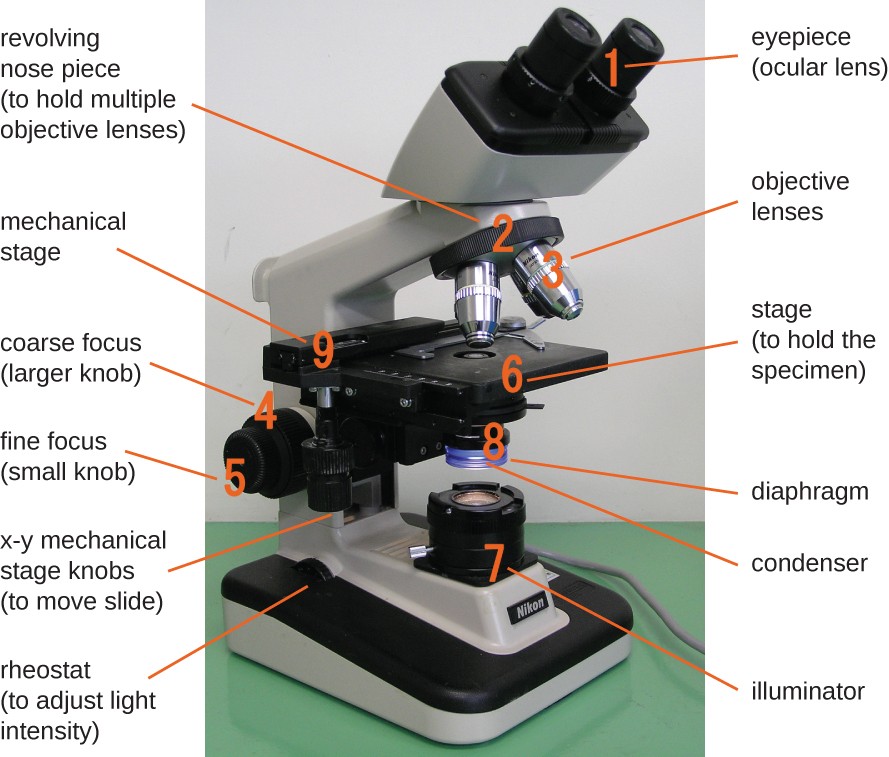
Parts of a microscope with functions and labeled diagram Q. List down the 18 parts of a Microscope. 1. Ocular Lens (Eye Piece) 2. Diopter Adjustment 3. Head 4. Nose Piece 5. Objective Lens 6. Arm (Carrying Handle) 7. Mechanical Stage 8. Stage Clip 9. Aperture 10. Diaphragm 11. Condenser 12. Coarse Adjustment 13. Fine Adjustment 14. Illuminator (Light Source) 15. Stage Controls 16. Base 17.
biology labeled microscope diagram - Clip Art Library
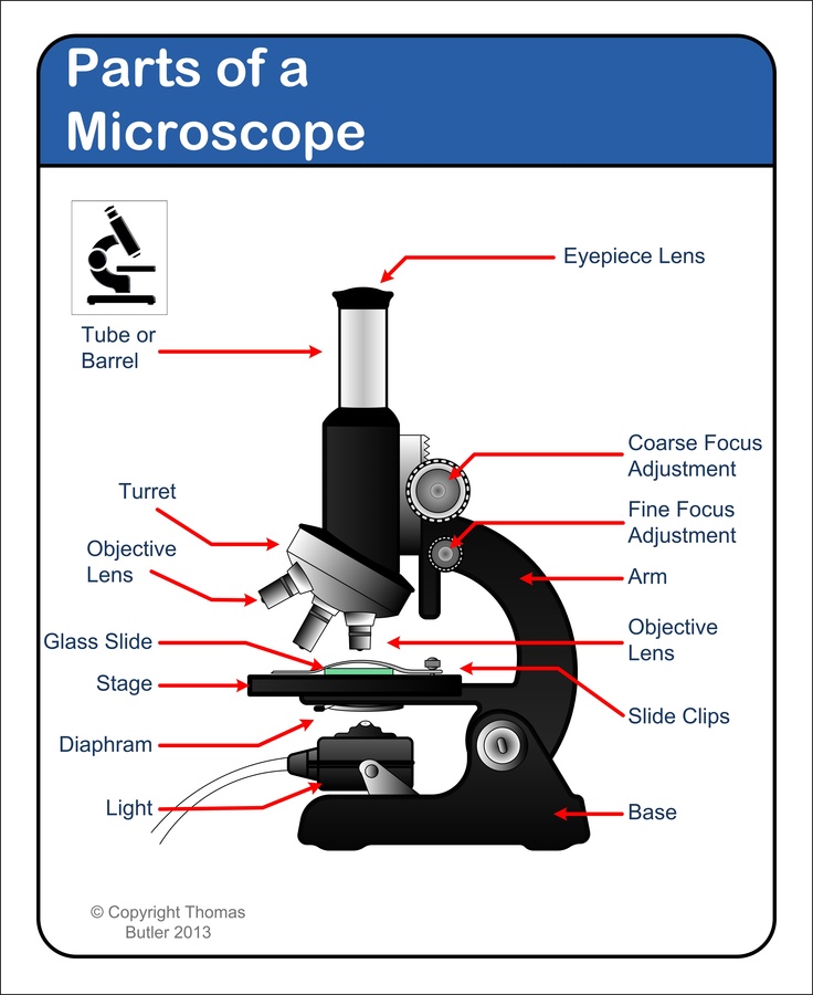
Label Microscope Diagram - EnchantedLearning.com Using the terms listed below, label the microscope diagram. arm - this attaches the eyepiece and body tube to the base. base - this supports the microscope. body tube - the tube that supports the eyepiece. coarse focus adjustment - a knob that makes large adjustments to the focus. diaphragm - an adjustable opening under the stage, allowing ...
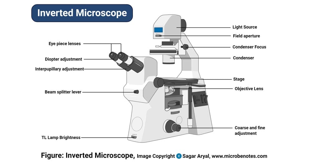
Inverted Microscope- Definition, Principle, Parts, Labeled ...
Labelled Diagram of Compound Microscope - Biology Discussion The below mentioned article provides a labelled diagram of compound microscope. Part # 1. The Stand: The stand is made up of a heavy foot which carries a curved inclinable limb or arm bearing the body tube. The foot is generally horse shoe-shaped structure (Fig. 2) which rests on table top or any other surface on which the microscope in kept.
Microscope- Definition, Parts, Functions, Types, Diagram, Uses
Compound Microscope Parts, Functions, and Labeled Diagram Compound Microscope Parts, Functions, and Labeled Diagram Parts of a Compound Microscope Each part of the compound microscope serves its own unique function, with each being important to the function of the scope as a whole.

Compound Microscope Parts
Microscope Parts, Function, & Labeled Diagram - slidingmotion Microscope parts labeled diagram gives us all the information about its parts and their position in the microscope. Microscope Parts Labeled Diagram The principle of the Microscope gives you an exact reason to use it. It works on the 3 principles. Magnification Resolving Power Numerical Aperture. Parts of Microscope Head Base Arm Eyepiece Lens

The Microscope
Experiment 1B | Lab01 | Virtual Edge| General Microbiology ...
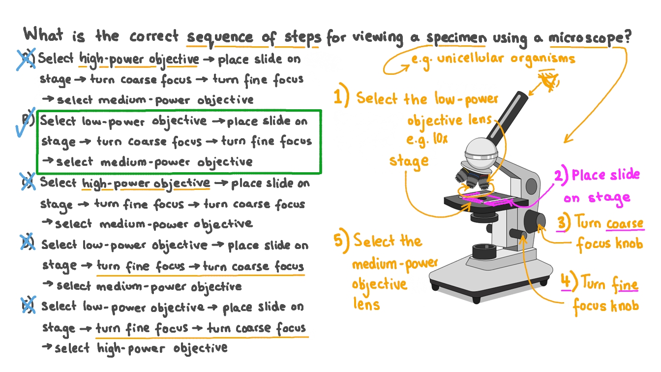
Stating the Correct Procedure for Viewing a Specimen under a Microscope
Junior cert Labelling Microscope - Labelled diagram

Cytology. Cytology. radiation used to illuminate the specimen ...

Compound Microscope Parts – Labeled Diagram and their ...

Microscope Diagram Labeled, Unlabeled and Blank | Parts of a ...

Parts of a Compound Microscope and Their Functions

A labeled diagram of a microscope. MLT 101. :) | Teaching ...
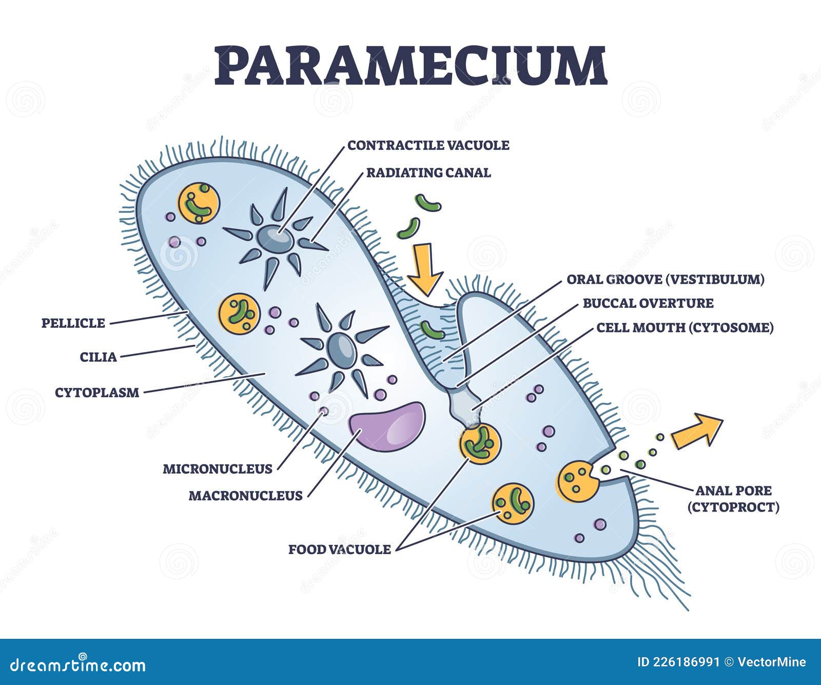
Paramecium Microscopic Closeup Structure with Anatomical ...

Microscope Terms Glossary | Earth science lessons, Biology ...

Diagram of Compound Microscope

Parts of a Microscope Labeling Activity

Microscope Labeled - yardlasopa
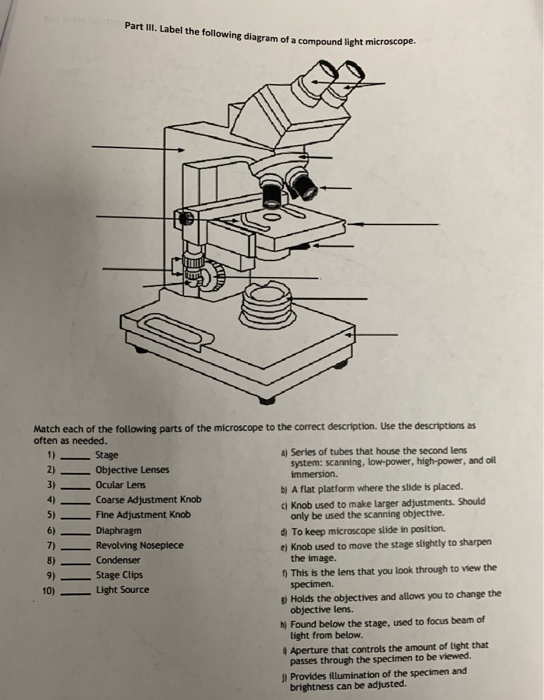
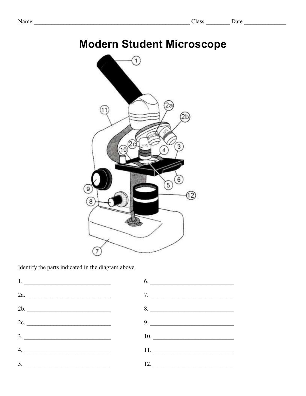
Solved Part III. Label the following diagram of a compound ...

Microscope Labeling Activity - SMART Board Activity - Interactive Review
Compound Microscope Parts, Functions, and Labeled Diagram ...
Microscope labeling, modern and classical types
Modified Science Diagram; Label Parts of a Microscope; Special Education

Compound Microscope Parts – Labeled Diagram and their ...

Compound Microscope Parts and Functions | Microscope parts ...

All Saints Online: Diagram for Labelling: Microscope
Microscope labeled diagram
Instruments of Microscopy | Microbiology | | Course Hero
Microscope Terminology
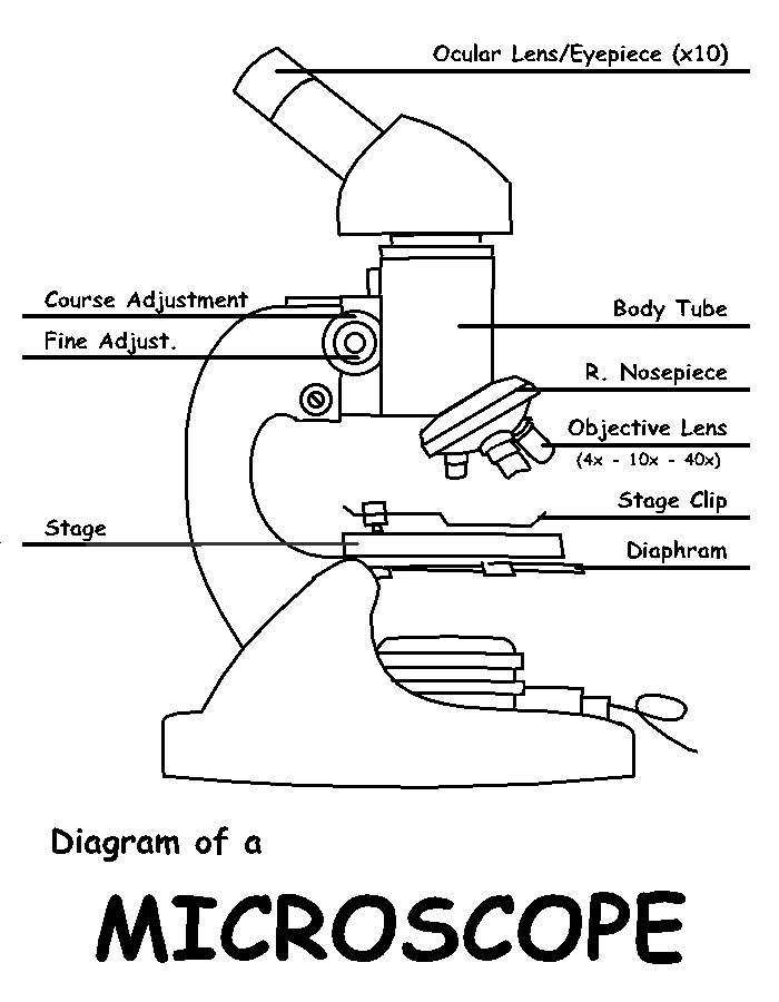
Diagram of a Microscope by ScienceDoodles on DeviantArt

Microscope, Microscope Parts, Labeled Diagram, and Functions
Microscope Diagram Parts - ClipArt Best

label microscope diagram | Charts | Microscope, Anatomy bones ...

Microscope Diagram - Label Diagram | Quizlet
Post a Comment for "44 diagram of a labeled microscope"