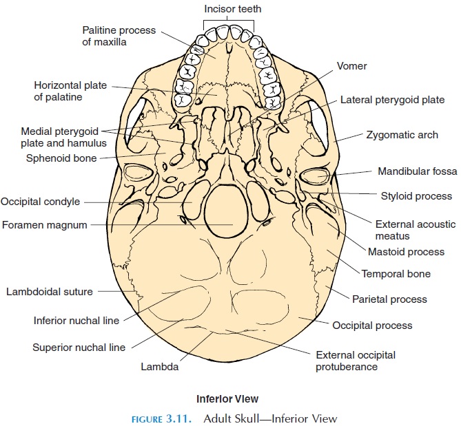
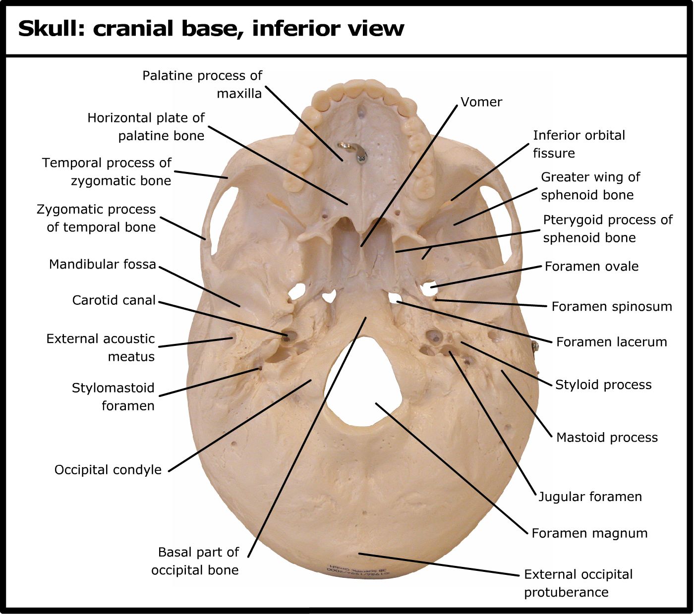
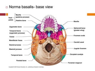
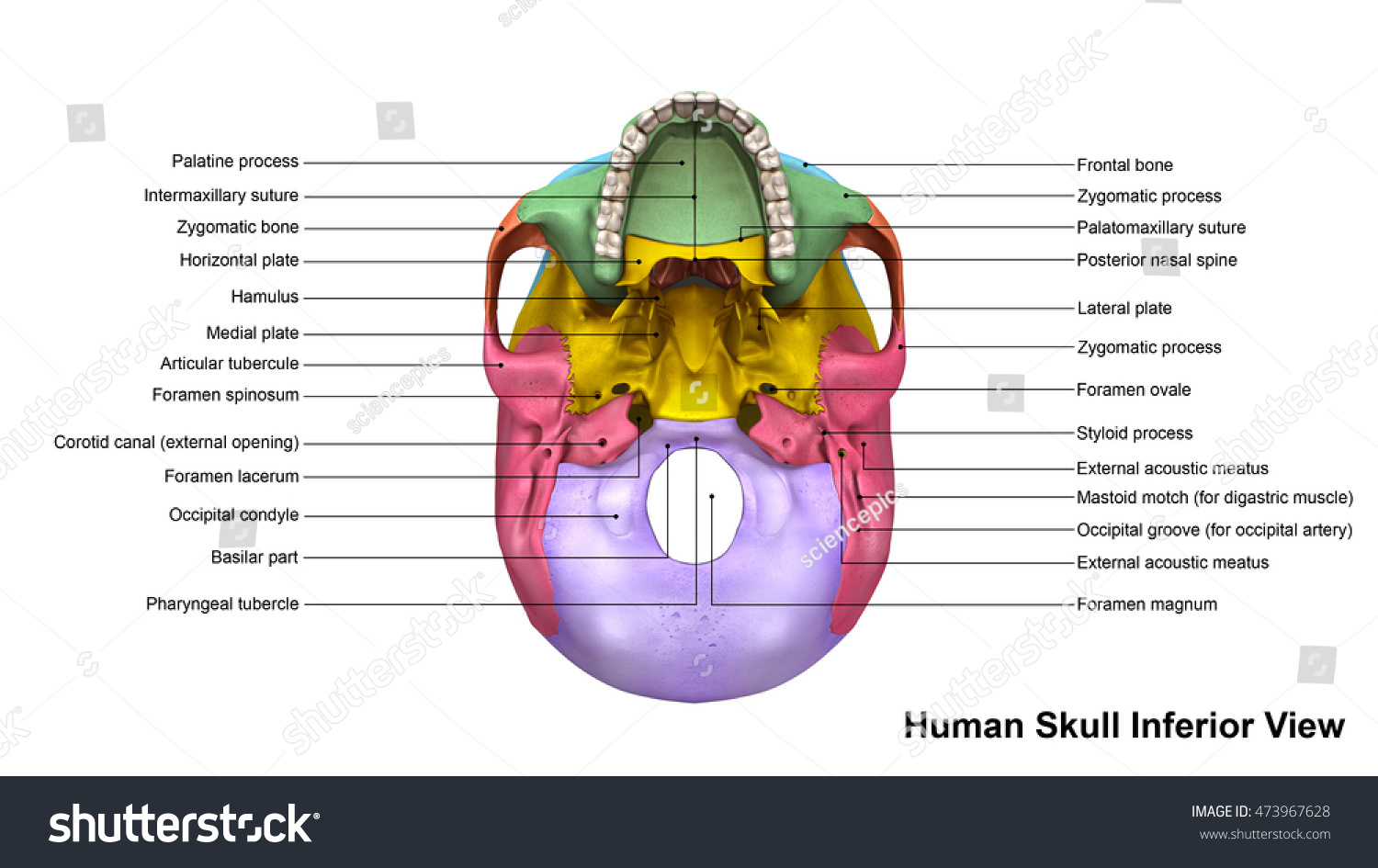
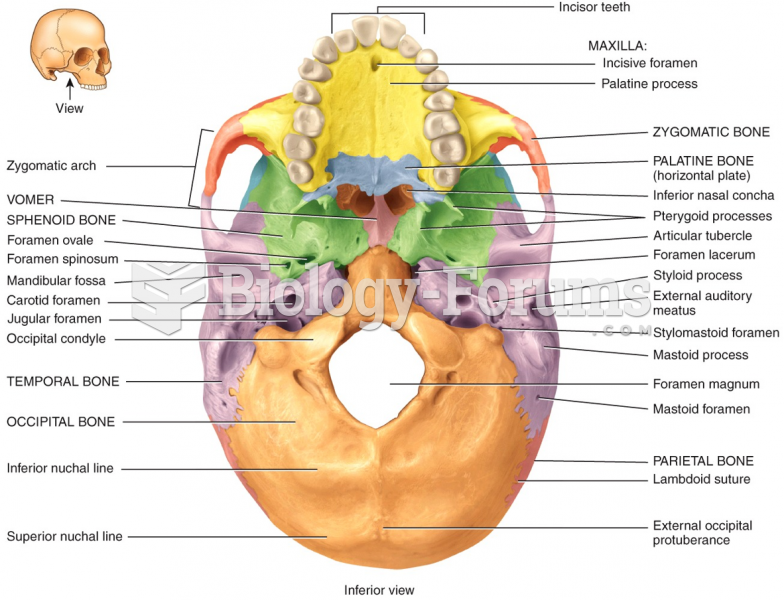
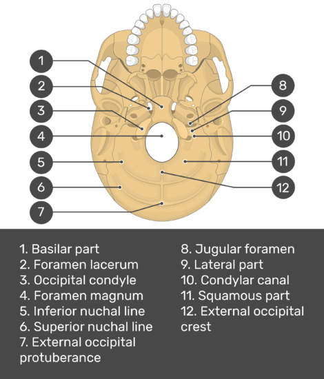
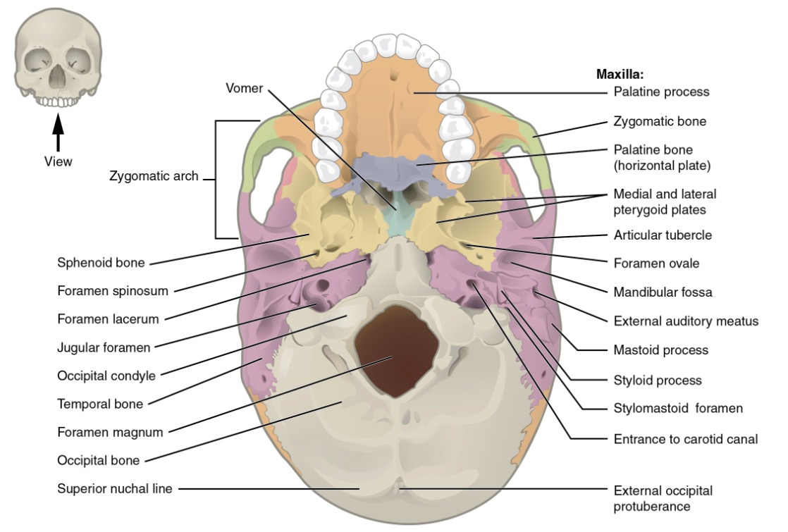
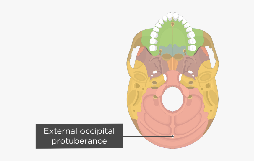
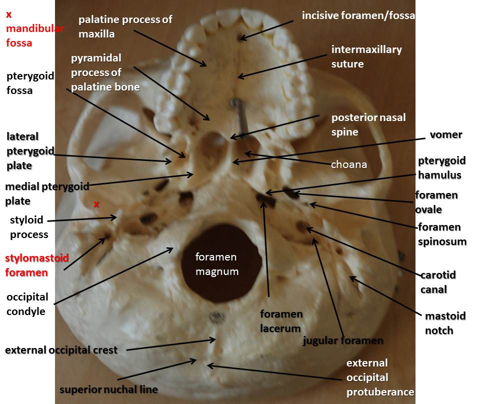
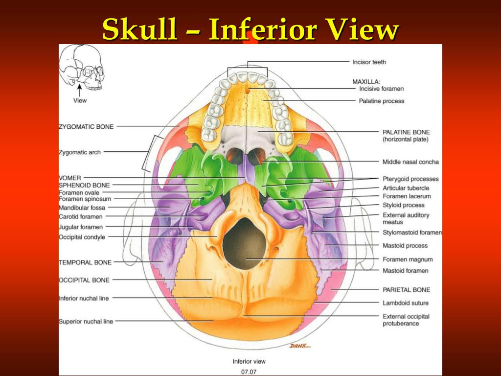
40 bones of the skull inferior view labeled
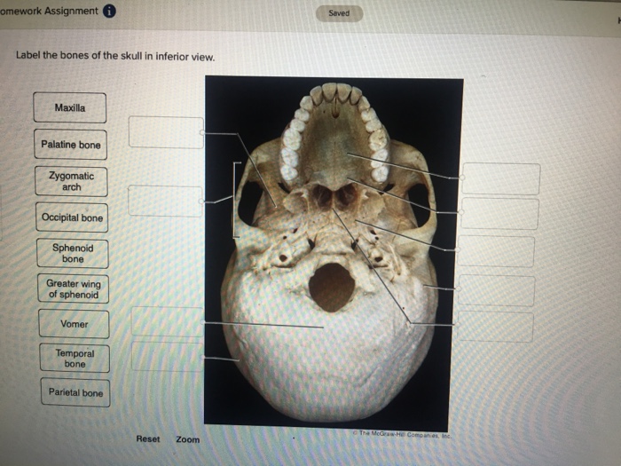
The Bones of the Skull | Human Anatomy and Physiology Lab (BSB 141 ... The bones that make up the cranium are called the cranial bones. The remainder of the bones in the skull are the facial bones. Figure 6.7 and Figure 6.8 show all the bones of the skull, as they appear from the outside. In Figure 6.9, some of the bones of the hard palate forming the roof of the mouth are visible because the mandible is not present. Saved omework Assignment Label the bones of the skull in inferior view ... A colored dot at the end of a leader line indicates a bone leader lines without a colored dot indicate bone markings. Note that vomer, sphenoid bone, and zygomatic bone will each be labeled twice. Key: 1. alveolar processes 2. carotid canal 3. ethmoid bone (perpendicular plate) 4. external occipital...
Anterior Skull Bones Anatomy - 2 osteology pocket dentistry, bones of ... Anterior Skull Bones Anatomy - 16 images - bones of the skull anterior view andrea kennedy flickr, bones of the skull, superior view of skull bones anatomy skeletal system pinterest, multi colored skull superior view of ethmoid and sphenoid flickr, ... Skull Inferior View Quiz, Authtool2.britishcouncil.org is an open platform for users to share ...
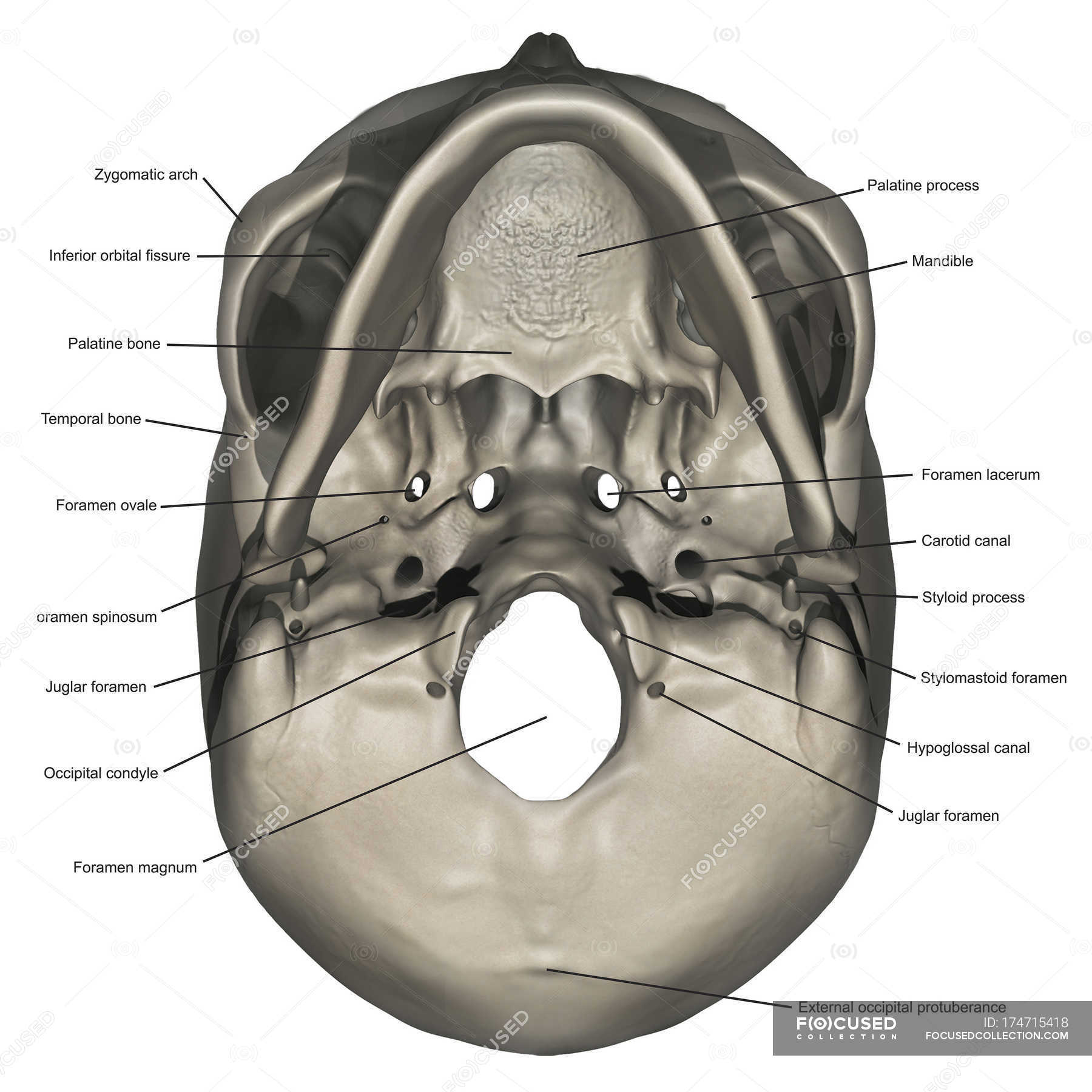
Bones of the skull inferior view labeled
The Skull | Anatomy and Physiology I - Lumen Learning A better view of the vomer bone is seen when looking into the posterior nasal cavity with an inferior view of the skull, where the vomer forms the full height of the nasal septum. The anterior nasal septum is formed by the septal cartilage , a flexible plate that fills in the gap between the perpendicular plate of the ethmoid and vomer bones. Greater palatine canal - Wikipedia Structure. The greater palatine canal starts on the inferior aspect of the pterygopalatine fossa.It goes through the maxilla and palatine bones to reach the palate, ending at the greater palatine foramen. From this canal, accessory canals branch off; these are known as the lesser palatine canals.. The canal is formed by a vertical groove on the posterior part of the maxillary surface … Scan atlas of anatomy of the face - e-Anatomy - IMAIOS 13.09.2021 · This head and neck anatomy atlas is an educational tool for studying the normal anatomy of the face based on a contrast enhanced multidetector computed tomography imaging (axial and coronal planes). Interactive labeled images allow a comprehensive study of the anatomical structures. Cross-sectional anatomy: CT of the head and neck
Bones of the skull inferior view labeled. Facial Bones of the Skull Mnemonic: Anatomy and Labeled Diagram - EZmed Nine = Nasal (2) Very = Vomer. Large = Lacrimal (2) Zucchini = Zygomatic (2) Pizzas = Palatine (2) The (2) denotes a pair, or 2, of those bones in the skull. We are now going to discuss the anatomy and important features of each facial bone in the order of the mnemonic. Image: The above mnemonic will help you remember the names of the facial bones. Anterior Skull Bones Anatomy - bones of the skull, rhinoplasty tutorial ... Anterior Skull Bones Anatomy - 16 images - bones of the skull posterior view flickr photo sharing, nasal bone anatomy borders function development kenhub, lateral view of the bones of the skull unlabeled, inferior jpeg image 1507 1093 pixels, anatomy bones of the skull Facial Bones Radiographic Anatomy - wikiRadiography. 8 Pics about Facial Bones Radiographic Anatomy - wikiRadiography : Mandible, lateral view with labels - Axial Skeleton Visual… | Flickr, Rotation of 3D skeleton.ribs,chest,anatomy,human,medical,body,skull and also Depressed skull fracture | Radiology Reference Article | Radiopaedia.org. Skeletal System • Anatomy & Function - GetBodySmart Skeletal System. The skeletal system is the body's structural framework. It mainly consists of bones, but also a network of ligaments, tendons and cartilage. The bones provide structure and protect the internal organs.. Cartilage is a flexible material found in joints that prevents bones from grinding against each other, which is important for joint movement.
Head and neck anatomy | Radiology Reference Article Head and neck anatomy is important when considering pathology affecting the same area. In radiology, the 'head and neck' refers to all the anatomical structures in this region excluding the central nervous system, that is, the brain and spinal cord and their associated vascular structures and encasing membranes i.e. the meninges. diagram of the skull labeled Inferior view of skull quiz. Skeleton axial skull diagram anterior labels anatomy visual bones atlas guide human skeletal system del flickr anatomía map read esqueleto photo. 17 Images about photo : Diagram Inferior View Of Skull Labeled - Diagram Media, Skull Diagram Side View - Diagram Media and also Skull inferior view - PurposeGames. Human Skull Anatomy Inferior View (Illustrations) - Human Bio Media The skull (cranium) is the skeletal structure of the head that supports the face and protects the brain. It is subdivided into the facial bones and the cranial bones. The facial bones underlie the facial structures, form the nasal cavity, enclose the eyeballs, and support the teeth of the upper and lower jaws. cranial bones labeled cranial bones labeled. Cranial nerves and swallowing: inferior view of cranial nerves exiting. Ethmoid bone anatomy plate bones skull perpendicular crista anterior medical dictionary physiology markings human nasal galli superior orbital articulation cribriform. Axial skeleton appendicular difference between vs study bone parts anatomy. cranial ...
SKULL inferior view - labeled This exhibit depicts the anatomy of the inferior skull including: the foramen magnum, occipital condyles, mastoid process, styloid process, mandibular fossa ... Skull Labeling - Inferior view Flashcards | Quizlet Start studying Skull Labeling - Inferior view. Learn vocabulary, terms, and more with flashcards, games, and other study tools. Search. Browse. Create. Log in Sign up. ... Human Anatomy & Physiology: Bones of the Skull 24 Terms. marissa_ann34. BIO 137 LAB EXAM 2 Skull 84 Terms. amber_richardson82. OTHER SETS BY THIS CREATOR. GOV FINAL 58 Terms. Learn skull anatomy with skull bones quizzes and diagrams 28.10.2021 · Overview image of an anterior view of the skull The idea behind using labeled diagrams is to get an overview of all of the structures within a given area. When it comes to testing your memory of these structures, previously having seen them altogether as a group should help you to remember them more easily. Wrist (lateral view) | Radiology Reference Article | Radiopaedia.org 19.09.2021 · The lateral wrist view is part of a three view series of the wrist and carpal bones. It is the orthogonal projection of the PA wrist. Indications The lateral wrist radiograph is requested for myriad reasons including but not limited to trauma, ...
Inferior Skull Labeling Quiz - PurposeGames.com An unregistered player played the game 2 weeks ago About this Quiz This is an online quiz called Inferior Skull Labeling There is a printable worksheet available for download here so you can take the quiz with pen and paper. From the quiz author Label the superficial markings and bones of the inferior skull. Your Skills & Rank Total Points 0
anterior skull bones skull inferior human carotid labeled anatomy bones canal diagram bone cranium physiology palatine foramen skeleton principles joints chapter internal process Body Parts Bones - Massage, Massage Videos, Massage Pictures, Massage parts bones body posterior massagenerd Anterior View Of The Skull Quiz
Inferior view of the base of the skull: Anatomy | Kenhub The parietal bones are difficult to visualise from the inferior view of the skull, however they can be seen articulating with the temporal and occipital bones. They form the posterosuperior part of the skull. Clinical points Young children who present with cleft palate have a failure of the two maxillae to unite in the midline.
Unlabeled Skull Diagram Inferior View of the Bones of the Skull Unlabeled Example - SmartDraw. Bone Diagrams: Skull (unlabeled) - large image. 1 of 1. diagram Diagram: The Respiratory System · Diagram: Human Heart · Bone Diagram: Human Skeleton. ... you will find Skull anatomy in it." "human anatomy bones skull 82 90 label the lateral features the skull - Human ...
structure of skull bones diagram skull bones unlabeled inferior blank diagram anatomy cranial diagrams holes foramen nerves Visible Interactive Ostrich - Labelled Head & Skull Anatomy - YouTube skull anatomy ostrich head labelled interactive The Skull -- Exploring Nature Educational Resource
Skull inferior view Quiz - PurposeGames.com This is an online quiz called Skull inferior view. There is a printable worksheet available for download here so you can take the quiz with pen and paper. From the quiz author. ... Bones of the Lower Appendicular Skeleton 10p Image Quiz. Spine 13p Image Quiz. Major Arteries of the systemic circulation 22p Image Quiz. Articulated shoulder ...
Solved Label the bones of the skull in inferior view. | Chegg.com Label the bones of the skull in inferior view. Greater wing of sphenoid Occipital bone Temporal bone Zygomatic arch Vomer Do Parietal bone Palatine bone < Prev 17 of 39 Next > Links search Bill Question: Label the bones of the skull in inferior view.
Solved Label the bones and bone features shown on the - Chegg Label the bones and bone features shown on the inferior view of the skull by clicking and dragging the labels to the correct location Styloid process Zygomatic arch Mandibular fossa External acoustic meatus Mastold process rences Palatine bone Temporal bone Zygomatic bone Ootipal condylo Foramen magnum
Anterior View of Skull - Sinclair Anterior View of Skull. Sphenoid. Mastoid Process of Temporal Bone. Temporal Bone. Sphenoid. Temporal Bone. Inferior Nasal Conchae . Ethmoid. 4 parts . Nasal Bones. Vomer. Frontal Bone . Zygomatic Bone. Maxilla (right) Mandible. Maxilla (left) PREVIOUS (LEFT ARROW) SHOW ALL (SPACEBAR) NEXT
Bones of the Skull- INFERIOR VIEW Diagram | Quizlet Start studying Bones of the Skull- INFERIOR VIEW. Learn vocabulary, terms, and more with flashcards, games, and other study tools.
Inferior Skull Bones & Features - MSU MediaSpace 5 Aug 2020 — Thumbnail for Inferior Skull Features. 01. Lindsey L... · Thumbnail for Inferior View of the Skull. 02. Occipital... · Thumbnail for Occipital ...
Petrous bone CT: normal anatomy| e-Anatomy - e-Anatomy 13.09.2021 · Full labeled CT Scan - Normal anatomy of the ear and petrous bone (temporal bone) ... allowed us to develop a functional and user-friendly atlas-based application for exploration of the temporal bone and skull base. ... axial slice wtith all structures labeled. Anatomy of the temporal bone: how to view the anatomical labels.
Bones of the Skull - Structure - Fractures - TeachMeAnatomy The cranium (also known as the neurocranium) is formed by the superior aspect of the skull. It encloses and protects the brain, meninges, and cerebral vasculature. Anatomically, the cranium can be subdivided into a roof and a base: Cranial roof - comprised of the frontal, occipital and two parietal bones. It is also known as the calvarium.
skull bone labeling Skull bones anatomy worksheets labeling head unlabeled face physiology human labeled system worksheet advanced printable coloring arm body muscle skeletal. Quizlet labeling markings ... Carlson Stock Art, Labeling the Adult Skull Bones Quiz and also Occipital Bone, Inferior View Quiz. Back Of Head Medical Term - Clip Art Library clipart-library ...
Facial Bones of the Skull Mnemonic: Anatomy and Labeled Diagram — EZmed 17.11.2020 · Use this facial bone mnemonic list to remember the anatomy, names, and structure of each of the facial bones of the skull. Uses labeled diagrams to show the structure and anatomy of each facial bone. Includes discussion about features and functions of the maxillary, mandible, nasal, conchae, zygomat
Inferior View of Bony Skull | Neuroanatomy | The Neurosurgical Atlas Zygomatic Arch. Inferior view of bony skull. The mandible has been removed to show the inferior surface and features of the skull in this specimen. Beginning anteriorly, the hard palate consists of the palatine processes of the maxillae and horizontal plates of the palatine bones. Just behind the anterior incisors is the incisive foramen.
Skull: Anatomy, structure, bones, quizzes | Kenhub 15.08.2022 · Skull (diagram, inferior view) The base of the skull extends from the superior nuchal lines of the occipital bones posteriorly to the upper incisors teeth anteriorly. This aspect of the skull contains a lot of important structures, including …
(Get Answer) - Label the bones of the skull in inferior view. Greater ... Label the bones of the skull in inferior view. Greater wing of sphenoid Occipital bone Temporal bone Zygomatic arch Vomer Do Parietal bone Palatine bone Links search Bill Expert's Answer Solution.pdf Next Previous Q: Q: Q: View Answer Q: Posted 7 months ago Recent Questions in Economics - Others Q:
Skull: Anatomy, structure, bones, quizzes | Kenhub The frontal bone is found superiorly while the mandible lies inferiorly, giving the skull an ovoid shape when looked at anteriorly. The frontal bone underlies the forehead; above the orbital cavities, the nasal bridge (which is formed jointly by the two nasal bones), and the frontal process of the zygomatic bone.
The Skull Bones Anatomy - Inferior View | GetBodySmart Let's start with taking a look at the cranial and facial bones from an anterior view before we dive into their markings from an inferior perspective. Facial Bones: Zygomatic bone ( os zygomaticum ). Maxilla bone ( os maxilla ). Palatine bone ( os palatinum ). Learn skull anatomy faster with these interactive skull bones quizzes and worksheets.
The Skull Bones - Orbital View | GetBodySmart The Skull Bones - Orbital View The Skull Bones - Orbital View Learn anatomy faster and remember everything you learn Start Now Bones Overview: Frontal bone ( os frontale ). Sphenoid bone ( os sphenoidale ). Ethmoid bone ( os ethmoidale ). Zygomatic bone ( os zygomaticum ). Lacrimal bone ( os lacrimale ). Maxilla bone ( os maxilla ). 1 2 3 4 5 6 7 8
Palatine process of maxilla - Wikipedia Base of skull. Inferior surface. Roof, floor, and lateral wall ... (Palatine process labeled at bottom right.) Medial surface of right maxilla. (Palatine process labeled at center ... Atlas image: rsa1p7 at the University of Michigan Health System – "Nasal septum, lateral view" "Anatomy diagram: 34257.000-1". Roche Lexicon
Skull - Inferior View - Human Body Help Skull - Inferior View Quiz yourself with the picture below. Scroll down for the answer key. Videos are at the bottom if you want to watch them first. Key: Incisive fossa Horizontal plate of the palatine bone Medial pterygoid plate Lateral pterygoid plate Foramen lacerum Foramen ovale Carotid canal Mandibular fossa Styloid process
Scan atlas of anatomy of the face - e-Anatomy - IMAIOS 13.09.2021 · This head and neck anatomy atlas is an educational tool for studying the normal anatomy of the face based on a contrast enhanced multidetector computed tomography imaging (axial and coronal planes). Interactive labeled images allow a comprehensive study of the anatomical structures. Cross-sectional anatomy: CT of the head and neck
Greater palatine canal - Wikipedia Structure. The greater palatine canal starts on the inferior aspect of the pterygopalatine fossa.It goes through the maxilla and palatine bones to reach the palate, ending at the greater palatine foramen. From this canal, accessory canals branch off; these are known as the lesser palatine canals.. The canal is formed by a vertical groove on the posterior part of the maxillary surface …
The Skull | Anatomy and Physiology I - Lumen Learning A better view of the vomer bone is seen when looking into the posterior nasal cavity with an inferior view of the skull, where the vomer forms the full height of the nasal septum. The anterior nasal septum is formed by the septal cartilage , a flexible plate that fills in the gap between the perpendicular plate of the ethmoid and vomer bones.



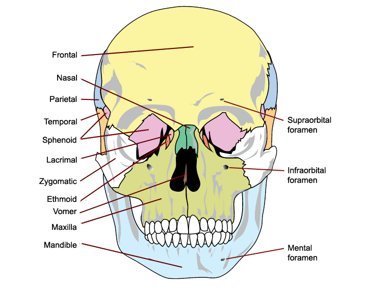
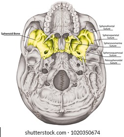








:background_color(FFFFFF):format(jpeg)/images/library/7562/inferior-base-of-the-skull_english.jpg)

Post a Comment for "40 bones of the skull inferior view labeled"