42 sarcomere labeled
Walter Herzog: H-index & Awards - Academic Profile - Research.com His study in Sarcomere is interdisciplinary in nature, drawing from both Myosin, Myofilament, Protein filament and Myofibril. His Cartilage study integrates concerns from other disciplines, such as Osteoarthritis, Articular cartilage, Composite material and Matrix. He most often published in these fields: Anatomy (35.80%) Sarc-Graph: Automated segmentation, tracking, and analysis of ... - PLOS As per the schematic, z-discs, which define the ends of an ≈ 2 μm sarcomere, are fluorescently labeled; (b) Movie still with a schematic illustration of deformation gradient F avg and tracked sarcomeres overlaid, sarcomere color corresponds to contraction level with red indicating the highest level of contraction; (c) Magnitudes of average ...
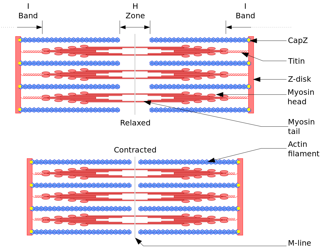
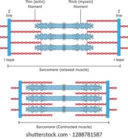
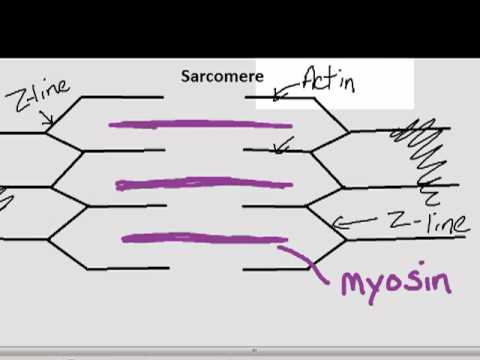
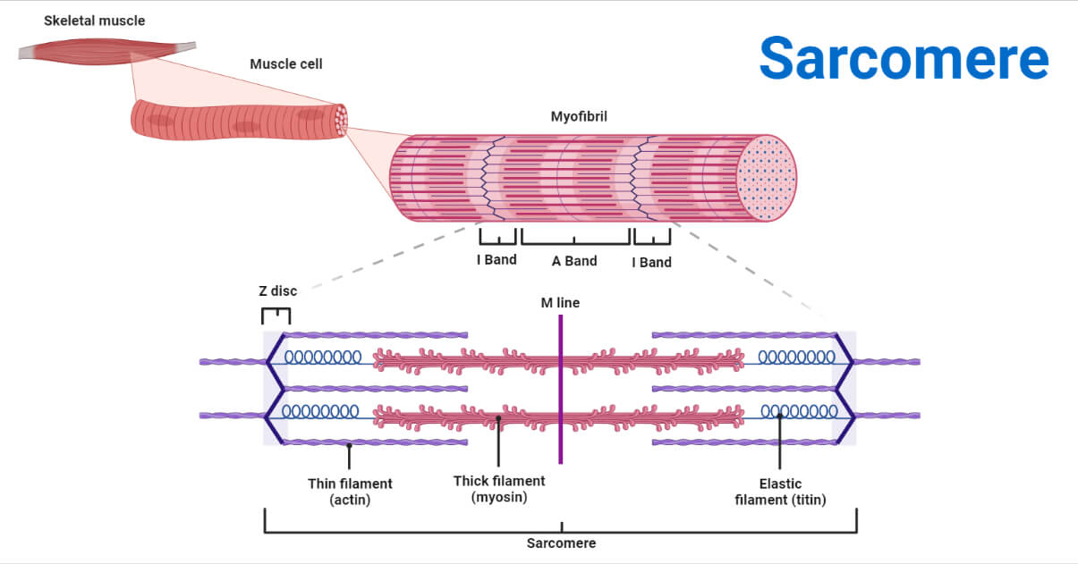
Sarcomere: anatomy, structure and function | Kenhub The sarcomere is the main contractile unit of muscle fiber in the skeletal muscle. Each sarcomere is composed of protein filaments ( myofilaments) that include mainly the thick filaments called myosin, and thin filaments called actin. The bundles of myofilaments are called myofibrils .
Sarcomere labeled
Physiology, Muscle Myocyte - StatPearls - NCBI Bookshelf The muscle cell, also known as the myocyte is the smallest subunit of all muscular tissues and organs throughout the body. It is here in the myocyte, where the physiological steps of muscle contraction and where the pathophysiology of numerous muscular diseases takes place. There are three types of muscle cells in the human body: skeletal, smooth, and cardiac muscle. The common function of ... Targeting the sarcomere in inherited cardiomyopathies - Nature The two main filaments in sarcomeres are the Ca 2+-dependent, regulatory thin filaments and the myosin-based, force-generating thick filaments 15 (Fig. 1).The thin filament is composed primarily ... Sarcomere Structure Labeled - muscular system, structure of the ... Sarcomere Structure Labeled. Here are a number of highest rated Sarcomere Structure Labeled pictures upon internet. We identified it from honorable source. Its submitted by management in the best field. We undertake this kind of Sarcomere Structure Labeled graphic could possibly be the most trending subject taking into account we allowance it ...
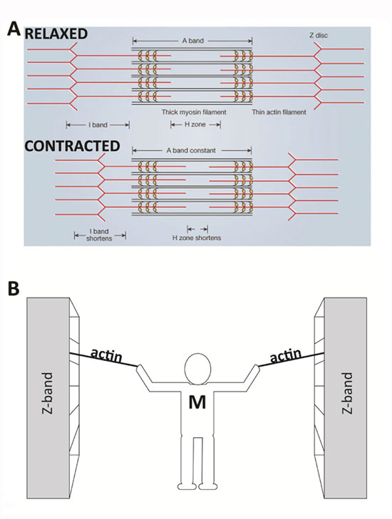
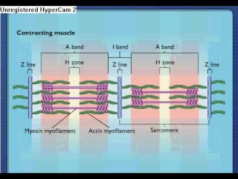
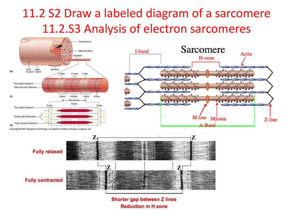
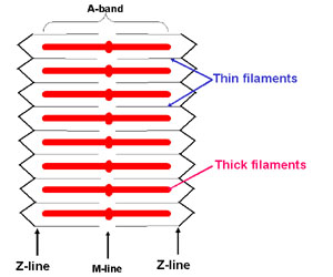
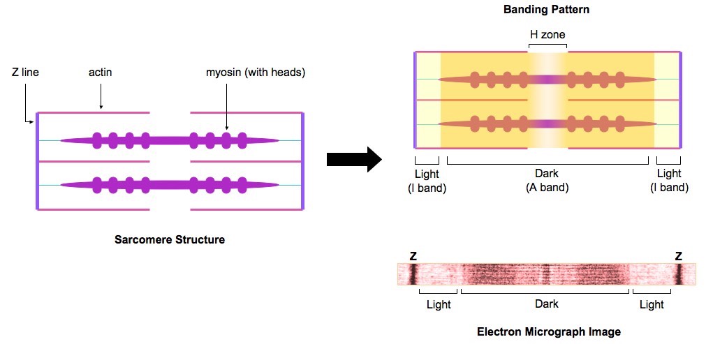
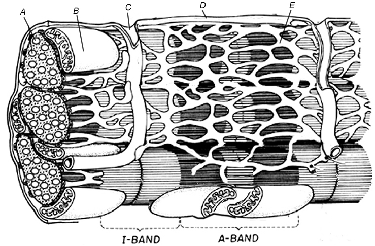
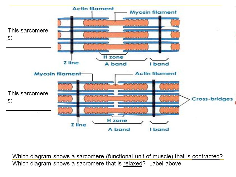
Sarcomere labeled. Length Tension Relationship - Human Physiology - 78 ... - 78 Steps Health Maximum relative tension (1.0 on the y axis) is achieved when the muscle is 100% to 120% of its resting length (sarcomere lengths from 2.0 to 2.25 |m). Increases or decreases in muscle (and sarcomere) lengths result in rapid decreases in tension. ... Human Anatomy and Physiology Study Course; Responses. kisanetHow to create maximum tension in ... Sarcomere protein modulation: The new frontier in cardiovascular ... Using blebbistatin we show that spin-labeled nucleotides bound to myosin have an oriented spectrum in the SRX in both slow and fast skeletal muscle. This is to our knowledge the first observation of oriented spin probes on the myosin motor domain in relaxed skeletal muscle fibers. ... In vivo sarcomere length (SL) measurements revealed that in ... In Figures 1 and 2 above, label the A-bands, I-bands, and ... - Brainly.com A sarcomere is composed of a thick and thin filaments, H-zone, A-, Z-, and I-bands.During contraction, the sarcomere gets shorter due to the shortening of the H-zone and the I-band.The other structures remain do not change. Sarcomere. In the sarcomere, we can identify the following components,. A-band.This band reflects the length of the thick filament, including a small portion of the thin ... Sarcomere Contraction - Glencoe.com Retirement: June 30, 2022 NOVELLA GLENCOE RETIREMENT: JUNE 30, 2022. On June 30, 2022, glencoe.mheducation.com and all of its associated sites will be retired and these sites will no longer be accessible. If you wish to retrieve any of the free resources available on glencoe.mheducation.com, please do so prior to June 30, 2022.
Thick-filer Muscle Contraction Model 1: Anatomy of a Sarcomere The ... Thick-filer Muscle Contraction Model 1: Anatomy of a Sarcomere The sarcomere is the functional (contractile) unit of skeletal muscle. It is the region of a myofibril between two Z discs. Each sarcomere is approximately 2 mm long. in tamen لال 1 Z M Z QUESTIONS 1. Label the thick horizontal filament THICK filament. 2. Numb and Numblike regulate sarcomere assembly and maintenance - JCI A sarcomere is the contractile unit of the myofibril in striated muscles such as cardiac and skeletal muscles. The assembly of sarcomeres depends on multiple molecules that serve as raw materials and participate in the assembly process. However, the mechanism of this critical assembly process remains largely unknown. The drawing and photomicrograph below show a relaxed sarcomere. Using ... The drawing and photomicrograph below show a relaxed sarcomere. Using the terms from ture indicated by a leader line or bracket. The number 2 in parentheses indicates that the struc rcomere. Using the terms from the key, identify each strue reses indicates that the structure will be labeled twice TETTY Key: a. actin filament b. A band c. H zone d. In silico identification of potential calcium dynamics and sarcomere ... For the sarcomere, boundary conditions and material properties we varied parameters in the range [50%, 150%], except for β 1 which we varied in the range [10%, 200%]. To cover both healthy and pathological calcium transient shapes, DCA, AMPL and TP parameters were varied in the range [10%, 200%], while for RT50 a smaller range was used ([10% ...
The Sarcomere and Sliding Filaments in Muscular Contraction: Definition ... While each sarcomere is small, several sarcomeres added together span the length of the muscle fiber. Each sarcomere consists of thick and thin bundles of proteins referred to as myofilaments. If... Label Sarcomere Quiz This is an online quiz called Label Sarcomere There is a printable worksheet available for download here so you can take the quiz with pen and paper. Your Skills & Rank Total Points 0 Get started! Today's Rank -- 0 Today 's Points One of us! Game Points 6 You need to get 100% to score the 6 points available Actions Add to favorites 0 favs Sarcomere Coloring Key This valuable student resource is intended for use in the undergraduate human anatomy and physiology class. The latest edition of Human Anatomy and Physiology Coloring Workbook is designed to help students learn introductory anatomy and physiology and is organized to complement the leading texts in the field. Virtually every structure of the ... Muscular Contraction: Cross-Bridge Formation - Study.com The sarcomere is the functional unit of striated muscle. Let's look at the cross-bridge within the context of a single sarcomere to understand how contraction occurs. ... UExcel Anatomy ...
File:Sarcomere.svg - Wikimedia Commons
Myofibril vs. Sarcomere - What's the difference? | Ask Difference A myofibril (also known as a muscle fibril or sarcostyle) is a basic rod-like organelle of a muscle cell. Muscles are composed of tubular cells called myocytes, known as muscle fibres in striated muscle, and these cells in turn contain many chains of myofibrils.
Sliding Filament Theory, Sarcomere, Muscle Contraction ...
Sarcomere Structure - striated muscle structure location function ... Sarcomere Structure. Here are a number of highest rated Sarcomere Structure pictures on internet. We identified it from honorable source. Its submitted by organization in the best field. We say you will this nice of Sarcomere Structure graphic could possibly be the most trending topic similar to we ration it in google help or facebook.
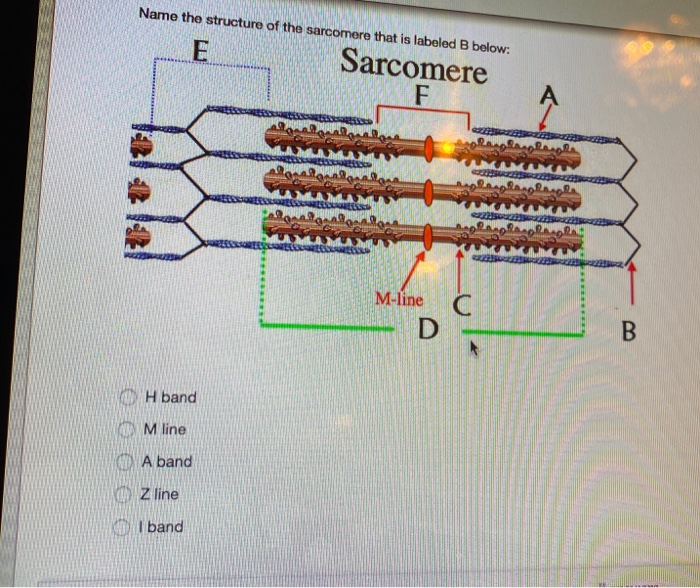
Solved Name the structure of the sarcomere that is labeled B ...
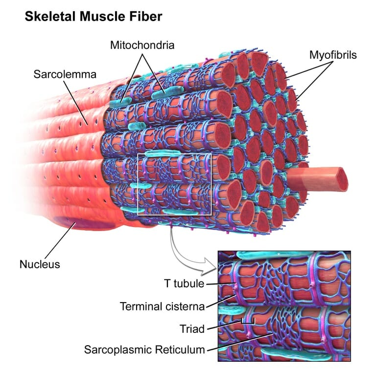
Muscle Growth | How Do Muscles Grow? - Anabolic.ca The sarcoplasm is the fluid area within the sarcolemma and between the myofibrils. The sarcoplasm is to muscle what the cytoplasm is to normal cells. It contains stored glycogen and oxygen-carrying myoglobin. Sarcomere Sarcomeres are the muscle's contractile units, and many are contained within one myofibril.
249 Sarcomere Images, Stock Photos & Vectors | Shutterstock
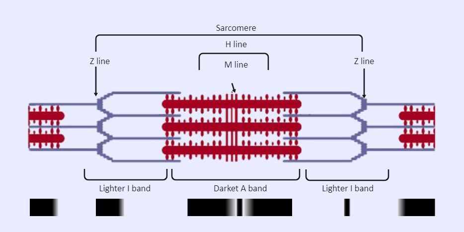
Sliding Filament - TeachMeAnatomy - Making Anatomy Simple The functional unit of contraction in a skeletal muscle fibre is the sarcomere, which runs from Z line to Z line. A sarcomere is broken down into a number of sections: Z line - where the actin filaments are anchored. M line - where the myosin filaments are anchored. I band - contains only actin filaments.

Sarcomere - an overview | ScienceDirect Topics
Alternative Splicing Mediated by RNA-Binding Protein RBM24 Facilitates ... Sarcomeres, segments spanning 2 Z-lines, are fundamental components of striated muscle tissue, and they orchestrate muscle contraction in both cardiac and skeletal muscle. Sarcomere formation is a complex process involving hundreds of genes and requiring precise assembly. However, the molecular mechanism underlying sarcomere assembly remains ...
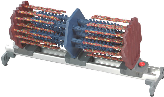
Sarcomere Model
Denoyer-Geppert® Sarcomere Model | Ward's Science - VWR The working sarcomere model helps bridge the link between cellular physiology and muscle tone, muscle cramps, knots and tears. Model also features a translucent sheath representing the sarcoplasmic reticulum, which can be wrapped around the sarcomere to offer greater understanding of membrane action potentials and the role calcium ion transport ...
Sarcomere Contraction - Process Of Muscle Contraction With Myosin & Actin
Skeletal muscle tissue: Histology | Kenhub The sarcomere is the functional unit of a skeletal muscle cell. Each sarcomere is about 2.5 micrometers in length. It is made up of multiple myosin and actin filaments oriented in parallel. The actin and myosin filaments overlap in certain places creating several bands and zones. A Z disc forms the boundary of the sarcomere on either side.
11.2 Movement. - ppt download
Art-labeling Activity: Sarcomere Structure Drag the labels...ask 3 Art-labeling Activity: Sarcomere Structure Drag the labels onto the diagram to identity structural features associated with a sarcomere. Reset Help A band bend Thinaments Mline Thick framanta Sarcomero Zine Tion Zone of overlap O Subna Request Answer. A horizontal demand curve or supply curve would be called: Group of answer choices Cross ...

sarcomere labeled diagram Diagram | Quizlet
DRUG DEVELOPMENT Copyright © 2022 Phenotypic screening with deep ... Loss of BAG3 in iPSC-CMs leads to sarcomere damage To develop a robust and reproducible loss-of-function model of DCM, we expressed small interfering RNAs (siRNAs) targeting BAG3 in human iPSC-CMs. First, using scramble (SCR) siRNA labeled with red fluorescent protein, we optimized conditions for iPSC-CM seeding
Muscle: The Histology Guide
Sarcomere Structure Labeled - muscular system, structure of the ... Sarcomere Structure Labeled. Here are a number of highest rated Sarcomere Structure Labeled pictures upon internet. We identified it from honorable source. Its submitted by management in the best field. We undertake this kind of Sarcomere Structure Labeled graphic could possibly be the most trending subject taking into account we allowance it ...
1. The sarcomere
Targeting the sarcomere in inherited cardiomyopathies - Nature The two main filaments in sarcomeres are the Ca 2+-dependent, regulatory thin filaments and the myosin-based, force-generating thick filaments 15 (Fig. 1).The thin filament is composed primarily ...
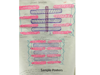
Sarcomere Diagram Poster Project by Anatomy Ready To Go | TpT
Physiology, Muscle Myocyte - StatPearls - NCBI Bookshelf The muscle cell, also known as the myocyte is the smallest subunit of all muscular tissues and organs throughout the body. It is here in the myocyte, where the physiological steps of muscle contraction and where the pathophysiology of numerous muscular diseases takes place. There are three types of muscle cells in the human body: skeletal, smooth, and cardiac muscle. The common function of ...
B1 Muscles and Movement | Brent Cornell
Sarcolemma Stock Photos, Stock Images and Vectors | Stockfresh
Sarcomere - Definition, Structure, Function and Quiz ...
Parts of the Sarcomere

10.2 Skeletal Muscle – Anatomy & Physiology

A schematic depicting the structure of an individual ...
Sarcomere (Muscle) Coloring

Identifying Regions in the Sarcomere
Muscles. Muscle Tissue Contains many mitochondria to power ...

Sarcomere Diagram | Quizlet
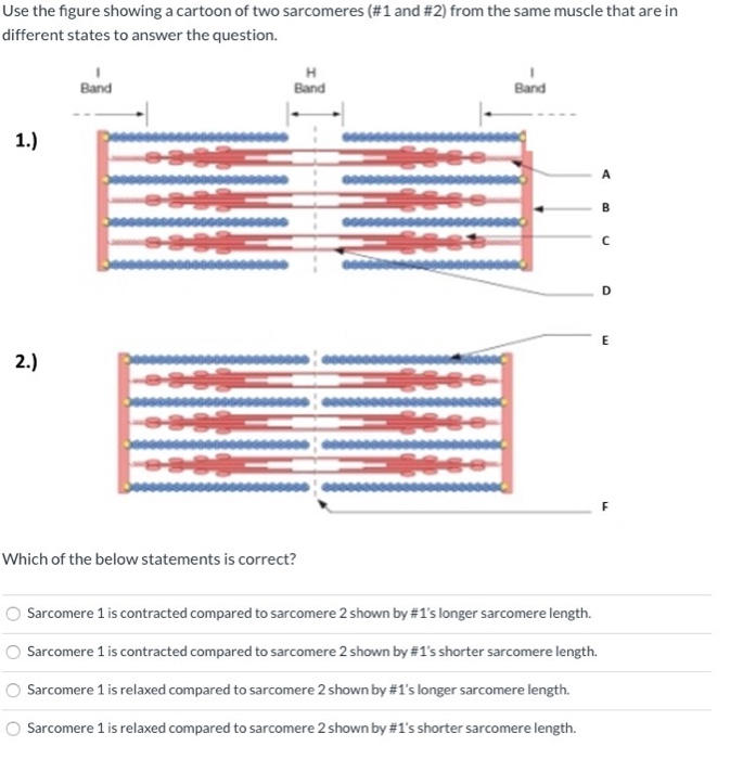
Solved 1.Which of the below statements is correct? 2.Which ...

Schematic diagram of a muscle sarcomere. The isotropic and ...
The Titin/Telethonin Complex
The Muscular System part 2 Muscle Physiology

Lesson Worksheet:Structure of Muscles | Nagwa

Sarcomere Labeling Quiz

View Image

Muscles and Movement AHL IB Biology. - ppt video online download
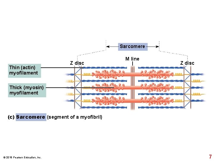
2018 Pearson Education Inc 1 2018 Pearson Education

Draw the diagram of a sarcomere of skeletal muscle showing ...

Untitled Document

10.2 Skeletal Muscle – Anatomy & Physiology
What is Muscle Contraction?- Definition and Mechanism

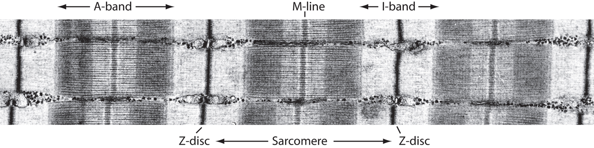
a Electron micrograph of a skeletal muscle sarcomere ...

Muscles | Baamboozle
Three distinct sarcomeric patterns of skeletal muscle ...

Sarcomere Structure - 11.2 Movement IB Biology HL Diagram ...

Sarcomere Structure Stock Illustrations – 45 Sarcomere ...
Sarcomere Labeled | EdrawMax Template
Sarcomere Model
Post a Comment for "42 sarcomere labeled"