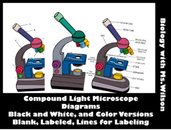
45 labelled diagram of compound microscope
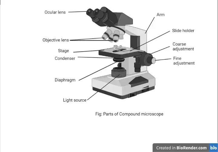
Compound microscope - their parts and function - Microscopy4kids Labeled diagram of a compound microscope. Optical components of a compound microscope. The term "compound" refers to the microscope having more than one lens. Compound microscopes generate magnified images through an aligned pair of the objective lens and the ocular lens. In contrast, "simple microscopes" have only one convex lens and ... Compound Microscope: Definition, Diagram, Parts, Uses, Working ... - BYJUS The compound microscope is mainly used for studying the structural details of cell, tissue, or sections of organs. The parts of a compound microscope can be classified into two: Non-optical parts Optical parts Non-optical parts Base The base is also known as the foot which is either U or horseshoe-shaped.
Parts of Stereo Microscope (Dissecting microscope) - labeled diagram ... If you would like to learn optical components of a compound microscope, please visit Compound Microscope Parts - Labeled Diagram and their Functions, and this article. How to use a stereo (dissecting) microscope. Follow these steps to put your stereo microscopes in work: 1. Set your microscope on a tabletop or other flat sturdy surface where ...
Labelled diagram of compound microscope
(i) Draw a neat labelled ray diagram of a compound microscope. Explain ... (i) Draw a neat labelled ray diagram of a compound microscope. Explain briefly its working. (ii) Why must both the objective and the eye-piece of a compound microscope have short focal lengths? cbse class-12 Share It On Facebook Twitter Email 1 Answer 0 votes answered May 15, 2018 by sanjaydas (89.7k points) selected May 24, 2018 by Vikash Kumar Compound Microscope: Parts of Compound Microscope - BYJUS Diagram Parts of the Compound Microscope Parts of Compound Microscope The parts of the compound microscope can be categorized into: Mechanical parts Optical parts (A) Mechanical Parts of a Compound Microscope 1. Foot or base It is a U-shaped structure and supports the entire weight of the compound microscope. 2. Pillar It is a vertical projection. Labelled Diagram of Compound Microscope The below mentioned article provides a labelled diagram of compound microscope. Part # 1. The Stand: The stand is made up of a heavy foot which carries a curved inclinable limb or arm bearing the body tube. The foot is generally horse shoe-shaped structure (Fig. 2) which rests on table top or any other surface on which the microscope in kept.
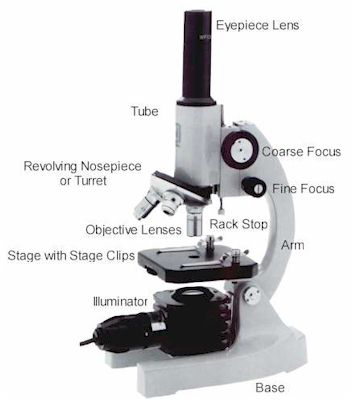
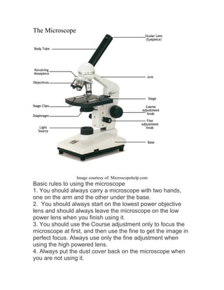
Labelled diagram of compound microscope. (a) Draw the labelled ray diagram for the formation of image by a ... (a) Draw the labelled ray diagram for the formation of image by a compound microscope. Derive an expression for its total magnification (or magnifying power), when the final image is formed at the near point. (b) Why both objective and eyepiece of a compound microscope must have short focal lengths? Parts of a microscope with functions and labeled diagram - Microbe Notes Parts of a microscope with functions and labeled diagram September 17, 2022 by Faith Mokobi Having been constructed in the 16th Century, Microscopes have revolutionalized science with their ability to magnify small objects such as microbial cells, producing images with definitive structures that are identifiable and characterizable. Microscope Parts and Functions Here are the important compound microscope parts... Eyepiece: The lens the viewer looks through to see the specimen. The eyepiece usually contains a 10X or 15X power lens. Diopter Adjustment: Useful as a means to change focus on one eyepiece so as to correct for any difference in vision between your two eyes. Microscope, Microscope Parts, Labeled Diagram, and Functions Revolving Nosepiece or Turret: Turret is the part of the microscope that holds two or multiple objective lenses and helps to rotate objective lenses and also helps to easily change power. Objective Lenses: Three are 3 or 4 objective lenses on a microscope. The objective lenses almost always consist of 4x, 10x, 40x and 100x powers. The most common eyepiece lens is 10x and when it coupled with ...
Microscope Parts, Function, & Labeled Diagram - slidingmotion Diaphragm. The diaphragm is also called as iris. This iris situates below the stage of the microscope. The function of the diaphragm is to control the amount of light that focuses on the specimen. This diaphragm can adjust the amount of light and intensity of light that falls on the specimen. In some standard and high-quality microscopes, this ... Compound Microscope Parts - Labeled Diagram and their Functions Labeled diagram of a compound microscope Major structural parts of a compound microscope Optical components of a compound microscope Eyepiece Eyepiece tube Objective lenses Nosepiece Specimen stage Coarse and fine focus knobs Rack stop Illuminator Condenser Abbe condenser Iris Diaphragm Condenser Focus Knob Summary An overview of microscopes Microscope: Parts Of A Microscope With Functions And Labeled Diagram. Figure: A diagram of a microscope's components. The microscope has three basic components: the head, the base, and the arm. Head:Occasionally, the head is considered the body. It holds the optical components of the upper part of the microscope. Base:The microscope's base provides great support. Compound Microscope - Types, Parts, Diagram, Functions and Uses It comes with a wide body and base. Its distinct parts include a condenser, illumination, focus lock, mechanical stage, and a revolving nosepiece which can hold up to five objectives. It usually has a binocular head, which makes long-term observation easy. Image 22: An example of a research compound microscope.
Compound Microscope Labeled Diagram | Quizlet Compound Microscope Labeled + − Flashcards Learn Test Match Created by meganplocher734 Terms in this set (14) Eyepiece/Ocular lens Contains the ocular lens Body tube A hollow cylinder that holds the eyepiece. Arm Part that supports the microscope. Stage Supports the slide or specimen Coarse adjustment Knob 16 Parts of a Compound Microscope: Diagrams and Video The 16 core parts of a compound microscope are: Head (Body) Arm Base Eyepiece Eyepiece tube Objective lenses Revolving Nosepiece (Turret) Rack stop Coarse adjustment knobs Fine adjustment knobs Stage Stage clips Aperture Illuminator Condenser Diaphragm Video: Parts of a compound Microscope with Diagram Explained Draw a labelled diagram of an image formed by a compound microscope ... Describe the compound microscope on the following headings (i) Labelled Ray diagram of formation of image. (ii) Magnifying power when final image is formed at least distance of distinct vision. Medium View solution > The final image formed by a compound microscope is : Easy View solution > View more More From Chapter Binocular Microscope Anatomy - Parts and Functions with a Labeled Diagram Now, I will describe all these non-optical parts of the light compound microscope with the labeled diagrams. The body tube of the microscope. The body tube is the solid support for the optical and mechanical parts of the microscope. There are two basic types of stand in the body tube of a light compound microscope - upright stand and inverted ...
Microscope Parts & Specifications | Microscope World Resources
A Study of the Microscope and its Functions With a Labeled Diagram ... A Study of the Microscope and its Functions With a Labeled Diagram To better understand the structure and function of a microscope, we need to take a look at the labeled microscope diagrams of the compound and electron microscope. These diagrams clearly explain the functioning of the microscopes along with their respective parts.

Compound Microscope Parts, Diagram Definition, Application ...
Diagram of a Compound Microscope - Biology Discussion Diagram of a Compound Microscope Article Shared by ADVERTISEMENTS: In this article we will discuss about:- 1. Essential Parts of Compound Microscope 2. Magnification of the Image of the Object by Compound Microscope 3. Resolution Power 4. Method for Studying Microbes 5. Measurement of the Size of Objects. Essential Parts of Compound Microscope:
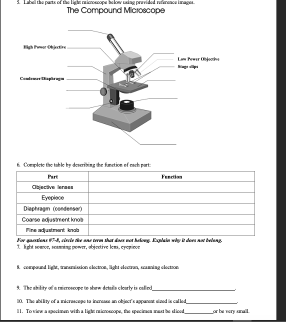
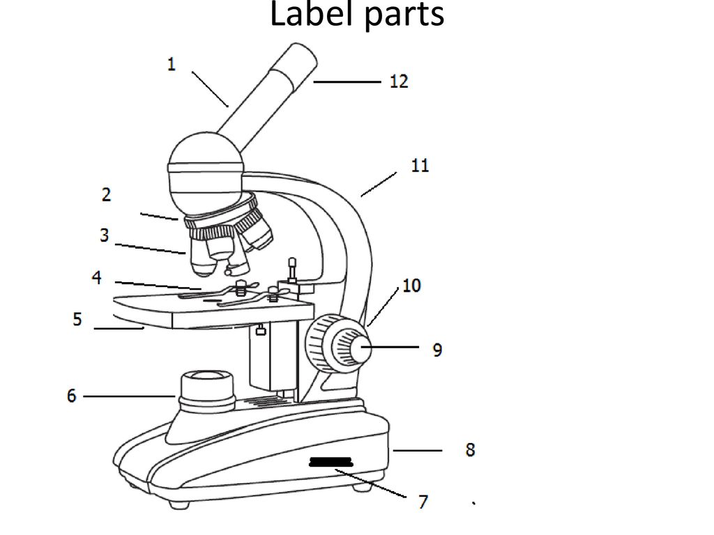
Solved 5. Label the parts of the light microscope below ...
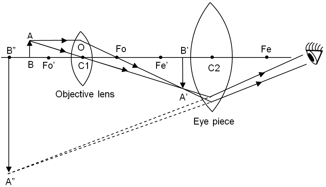
(a) Draw a labelled ray diagram of a compound microscope. (b) Derive an ... (a) Labelled diagram of compound microscope. The objective lens form image A' B' near the first focal point ofeyepiece. (b) Angular magnification of objective lens m0 = linear magnification h'/h where L is the distance between second focal point of the objective and first focal point of eyepiece.

Label diagram of compound microscope - Science - The ...
Compound Microscope- Definition, Labeled Diagram, Principle, Parts, Uses Parts of a Compound Microscope Eyepiece And Body Tube. The eyepiece is the lens through which the viewer looks to see the specimen. It usually contains a 10X or 15X power lens. The body tube connects the eyepiece to the objective lenses. Objectives and Stage Clips Objective Lenses are one of the most important parts of a Compound Microscope.

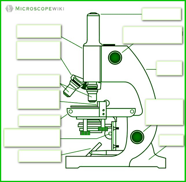
Microscope Diagram Labeled, Unlabeled and Blank | Parts of a ...
Labelled Diagram of Compound Microscope The below mentioned article provides a labelled diagram of compound microscope. Part # 1. The Stand: The stand is made up of a heavy foot which carries a curved inclinable limb or arm bearing the body tube. The foot is generally horse shoe-shaped structure (Fig. 2) which rests on table top or any other surface on which the microscope in kept.

Parts of a microscope with functions and labeled diagram
Compound Microscope: Parts of Compound Microscope - BYJUS Diagram Parts of the Compound Microscope Parts of Compound Microscope The parts of the compound microscope can be categorized into: Mechanical parts Optical parts (A) Mechanical Parts of a Compound Microscope 1. Foot or base It is a U-shaped structure and supports the entire weight of the compound microscope. 2. Pillar It is a vertical projection.
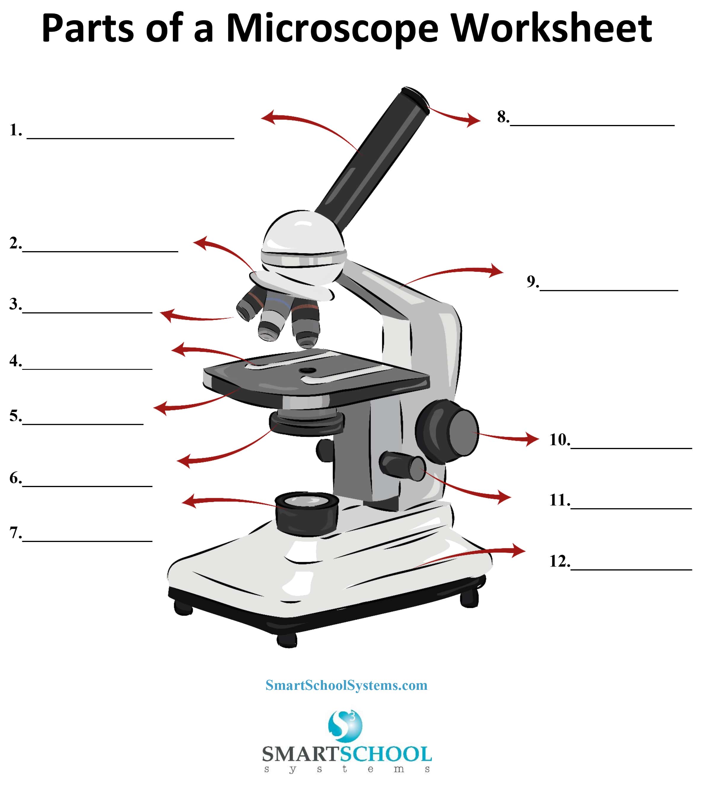
Parts of a Microscope - SmartSchool Systems
(i) Draw a neat labelled ray diagram of a compound microscope. Explain ... (i) Draw a neat labelled ray diagram of a compound microscope. Explain briefly its working. (ii) Why must both the objective and the eye-piece of a compound microscope have short focal lengths? cbse class-12 Share It On Facebook Twitter Email 1 Answer 0 votes answered May 15, 2018 by sanjaydas (89.7k points) selected May 24, 2018 by Vikash Kumar

Simple Microscope Definition, Magnification, Parts And Uses
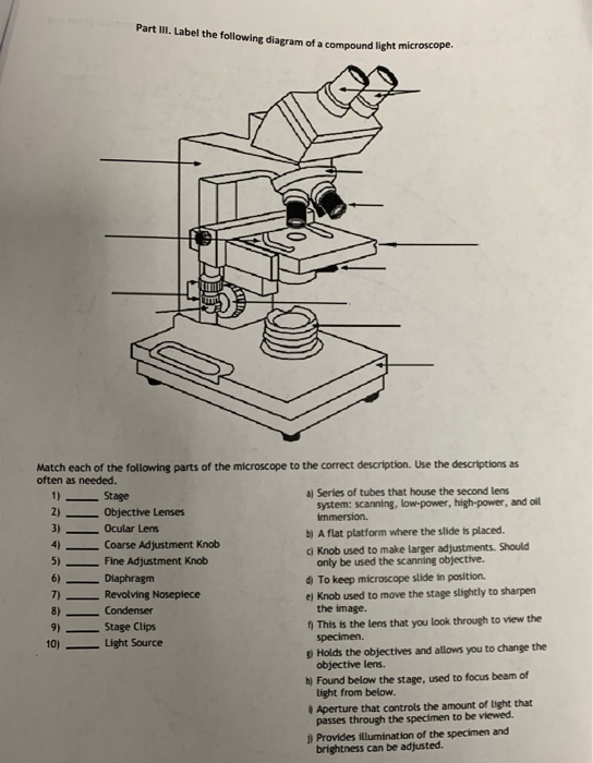
Solved Part III. Label the following diagram of a compound ...
Draw a labelled ray diagram of a compound microscope and ...
Compound Microscope Review - ppt download

Parts of a Microscope with Their Functions – Microbe Online
Parts of a Microscope with Their Functions – Microbe Online

Labelled Diagram of Compound Microscope | Figure Of Compound ...

Draw a labelled diagram of an image formed by a compound ...
i) Draw a neat labelled ray diagram of a compound microscope ...

Label the Microscope Diagram | Download Scientific Diagram
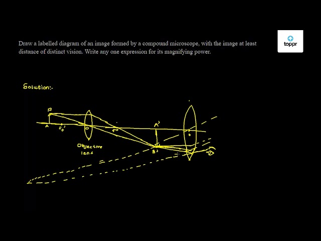
Draw a labelled diagram of an image formed by a compound microscope, with the image at least distance of distinct vision. Write any one expression for its magnifying power.

Compound Microscope Parts – Labeled Diagram and their ...

Compound Microscope Parts and Functions | Microscope parts ...

Compound Microscope Parts, Functions, and Labeled Diagram ...
Label The Parts Of A Compound Microscope Teaching Resources | TPT

Label the microscope — Science Learning Hub
Microscope labeled diagram

What is a Compound Microscope? | Microscope World Blog

Draw a labelled ray diagram of compound microscope and derive ...

Compound Microscope Parts – Labeled Diagram and their ...
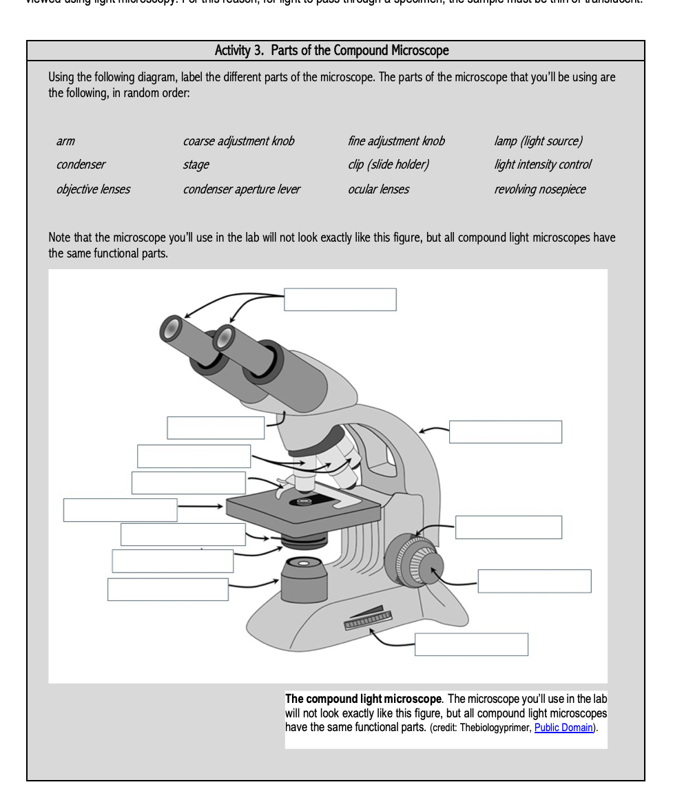
Solved Activity 3. Parts of the Compound Microscope Using ...

Can someone can send me diagram of this compound microscope ...

This is a common compound microscope. Label its parts from A ...

Microscope labeled diagram

Compound Light Microscope Labeling Diagram | Quizlet

Compound Microscope – Diagram (Parts labelled), Principle and ...

Living Environment Course

Compound microscope - their parts and function - Microscopy4kids

Compound Microscope Parts, Functions, and Labeled Diagram ...
The Compound Light Microscope Label the following parts on ...

Microscope Parts and Functions
i) Draw a neat labelled ray diagram of a compound microscope ...

Describe the structure of compound microscope with well ...

Compound Microscope Labeled Diagram | Quizlet
Simple Microscope - Diagram (Parts labelled), Principle ...

Microscope Labeling
Microscopy- History, Classification, Terms, Diagram

Draw a labelled diagram of a compound microscope.
Post a Comment for "45 labelled diagram of compound microscope"