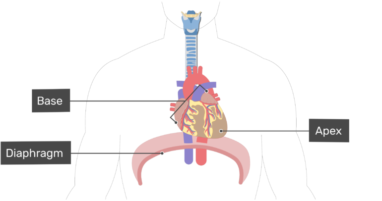
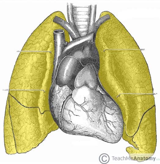
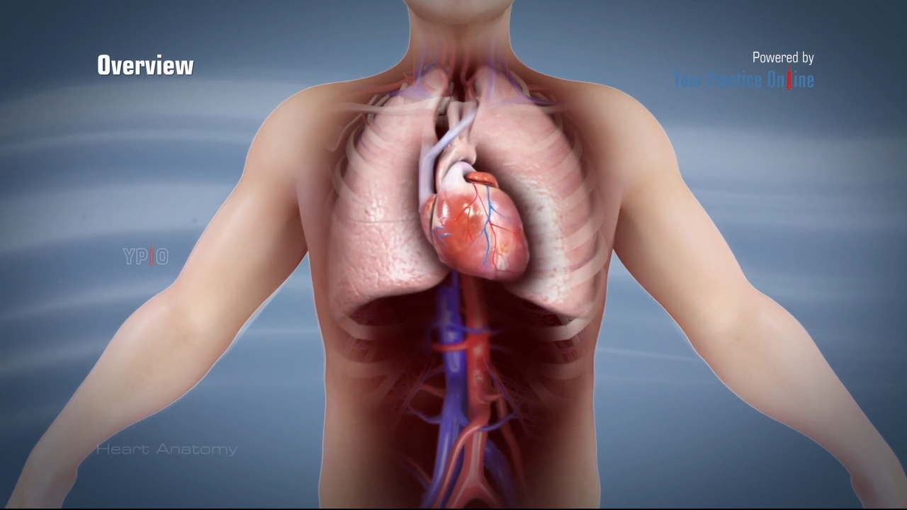
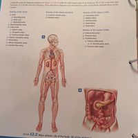
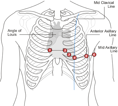
43 correctly label the following anatomical features of the heart and thoracic cage.
The Thoracic Cage | Anatomy and Physiology I - Lumen Learning The sternum is the elongated bony structure that anchors the anterior thoracic cage. It consists of three parts: the manubrium, body, and xiphoid process. The manubrium is the wider, superior portion of the sternum. The top of the manubrium has a shallow, U-shaped border called the jugular (suprasternal) notch. This can be easily felt at the anterior base of the neck, between the medial ends of the clavicles. Ch. 19 Circulatory System- heart Flashcards | Quizlet Correctly label the following internal anatomy of the heart. Drag each label to the location of each structure described. Explanation The heart functions to first pump deoxygenated blood returning from the body to the lungs in order to release carbon dioxide and reoxygenate the blood.
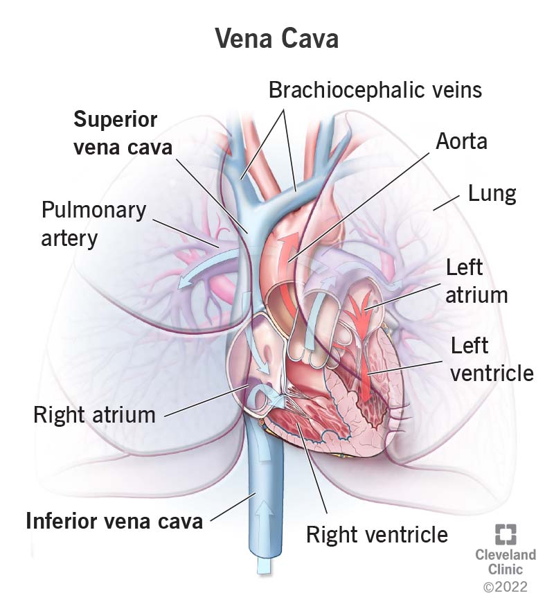
Question : correctly label the following anatomical features of the ... Answer: 1: Right atrium: It receives deoxygenated blood from superior and inferior venacava. 2: Right lung: Right lung has three lobes and along wit …. View the full answer. Transcribed image text: Correctly label the following anatomical features of the heart and thoracic cage. Right atrium Right lung Superior vena cava Left atrium Aortic arch Right ventricle Apex of heart Diaphragm.

Correctly label the following anatomical features of the heart and thoracic cage.
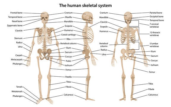
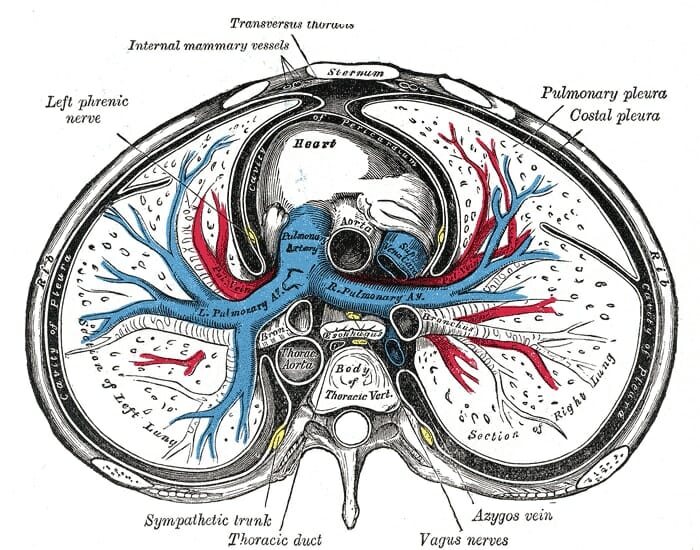
Thoracic cage: Anatomy and clinical notes | Kenhub The thoracic cage takes the form of a domed bird cage with the horizontal bars formed by ribs and costal cartilages. It is supported by the vertical sternum (anteriorly) and the 12 thoracic vertebrae (posteriorly). The main function of the thoracic cage is to support thorax and protect the vital structures within it (e.g. heart, lungs, aorta, etc). Anatomy & Physiology: The Unity of Form and Function - Quizlet Correctly label the following anatomical features of the heart and thoracic cage. Correctly label the following structures related to the position of the heart in the thorax. Correctly label the following anatomical features of the thoracic cavity. Correctly label the following parts of the pericardium and the heart walls. Thoracic cavity | Description, Anatomy, & Physiology | Britannica It contains the lungs, the middle and lower airways—the tracheobronchial tree—the heart, the vessels transporting blood between the heart and the lungs, the great arteries bringing blood from the heart out into general circulation, and the major veins into which the blood is collected for transport back to the heart. The heart is covered by a fibrous membrane sac called the pericardium that blends with the trunks of the vessels running to and from the heart.
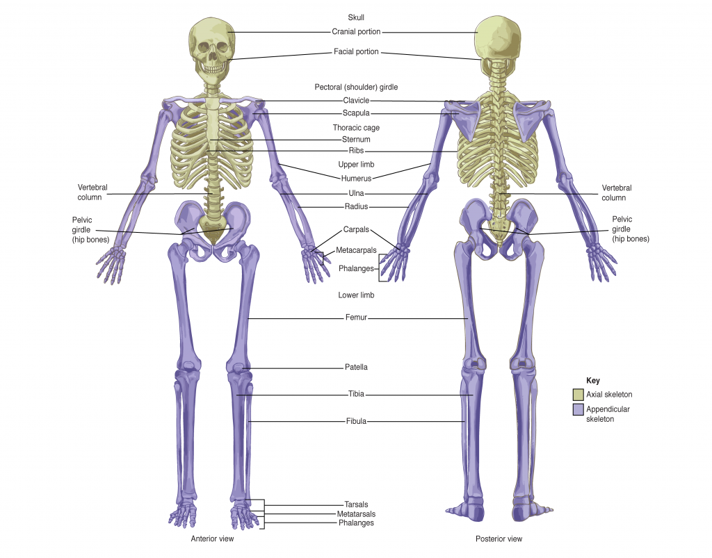
Correctly label the following anatomical features of the heart and thoracic cage.. 7.5 The Thoracic Cage - Anatomy & Physiology The thoracic cage (rib cage) forms the thorax (chest) portion of the body. It consists of the 12 pairs of ribs with their costal cartilages and the sternum (Figure 7.5.1). The ribs are anchored posteriorly to the 12 thoracic vertebrae (T1-T12). The thoracic cage protects the heart and lungs. Thorax: Anatomy, wall, cavity, organs & neurovasculature | Kenhub The thoracic, or chest wall, consists of a skeletal framework, fascia, muscles, and neurovasculature - all connected together to form a strong and protective yet flexible cage. The thorax has two major openings: the superior thoracic aperture found superiorly and the inferior thoracic aperture located inferiorly. The superior thoracic aperture opens towards the neck. The Thoracic Cage · Anatomy and Physiology The ribs are anchored posteriorly to the 12 thoracic vertebrae (T1-T12). The thoracic cage protects the heart and lungs. Sternum. The sternum is the elongated bony structure that anchors the anterior thoracic cage. It consists of three parts: the manubrium, body, and xiphoid process. The manubrium is the wider, superior portion of the sternum. Solved Correctly label the following anatomical features of - Chegg Question: Correctly label the following anatomical features of the heart and thoracic cage. Rectus abdominis 4th rib Diaphragm INC Sternum UD OULU 3rd rib ( 1 This problem has been solved! You'll get a detailed solution from a subject matter expert that helps you learn core concepts. See Answer Show transcribed image text Expert Answer
Thoracic cavity | Description, Anatomy, & Physiology | Britannica It contains the lungs, the middle and lower airways—the tracheobronchial tree—the heart, the vessels transporting blood between the heart and the lungs, the great arteries bringing blood from the heart out into general circulation, and the major veins into which the blood is collected for transport back to the heart. The heart is covered by a fibrous membrane sac called the pericardium that blends with the trunks of the vessels running to and from the heart. Anatomy & Physiology: The Unity of Form and Function - Quizlet Correctly label the following anatomical features of the heart and thoracic cage. Correctly label the following structures related to the position of the heart in the thorax. Correctly label the following anatomical features of the thoracic cavity. Correctly label the following parts of the pericardium and the heart walls. Thoracic cage: Anatomy and clinical notes | Kenhub The thoracic cage takes the form of a domed bird cage with the horizontal bars formed by ribs and costal cartilages. It is supported by the vertical sternum (anteriorly) and the 12 thoracic vertebrae (posteriorly). The main function of the thoracic cage is to support thorax and protect the vital structures within it (e.g. heart, lungs, aorta, etc).







:max_bytes(150000):strip_icc()/2313_The_Lung_Pleurea-6c90e267b8c9452289ab976ce32d1b83.jpg)







:background_color(FFFFFF):format(jpeg)/images/library/11130/Thorax_Thumbnail_02.png)


Post a Comment for "43 correctly label the following anatomical features of the heart and thoracic cage."